 뒤로
Innate Defenses and Inflammation: The Immune System in Anatomy & Physiology
스터디 가이드 - 스마트 노트
 자료에 맞춘 맞춤형 노트, 핵심 정의, 예시, 맥락을 확장해 제공합니다.
The Immune System: Overview
Introduction to the Immune System
The immune system is a functional system that provides resistance to disease and is essential for maintaining health. It is composed of two intrinsic systems: the innate (nonspecific) defense system and the adaptive (specific) defense system.
Innate Defense System: Provides the first and second lines of defense against pathogens.
Adaptive Defense System: Provides the third line of defense, targeting specific foreign substances.
Multifunctional Roles of Blood and Immune System
Blood and the immune system work together to protect the body from infection, dispose of debris, and facilitate repair.
Blood: Transports immune cells and mediators throughout the body.
Immune System: Detects and eliminates pathogens, and initiates healing processes.
Innate (Nonspecific) Defenses
First Line of Defense: Surface Barriers
The first line of defense consists of physical and chemical barriers that prevent the entry of pathogens.
Physical Barriers: Skin and mucous membranes act as mechanical barriers.
Chemical Barriers: Include acidity (acid mantle), enzymes (lysozyme in saliva, mucus, and lacrimal fluid), mucin, defensins, and other antimicrobial chemicals.
Keratin: Provides resistance to acids, bases, bacterial enzymes, and toxins.
Mucosae: Trap and remove microorganisms.
Example: Acid Mantle
The acidity of skin and vaginal secretions inhibits microbial growth, forming a protective acid mantle.
Example: Lysozyme
Lysozyme in saliva and tears destroys bacteria by breaking down their cell walls.
Example: Mucin
Sticky mucus traps pathogens in the digestive and respiratory tracts.
Example: Defensins
Antimicrobial peptides that disrupt microbial membranes.
Example: Sebum and Dermicidin
Lipids in sebum and dermicidin in sweat are toxic to some bacteria.
Respiratory System Modifications
Cilia and mucus in the respiratory tract help remove pathogens.
Second Line of Defense: Cells and Chemicals
When surface barriers are breached, the second line of defense is activated. This includes phagocytes, natural killer (NK) cells, inflammation, antimicrobial proteins, and fever.
Phagocytes: Chief cells include macrophages and neutrophils.
Pattern Recognition Receptors: Allow cells to recognize and bind to microbial structures.
Phagocytosis: Mechanism and Steps
Phagocytes and Their Types
Macrophages: Develop from monocytes; chief phagocytic cells.
Free Macrophages: Wander through tissues (e.g., alveolar macrophages).
Fixed Macrophages: Permanent residents in organs (e.g., stellate macrophages in liver, microglia in brain).
Steps of Phagocytosis
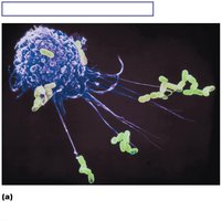
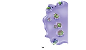
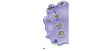
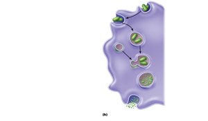
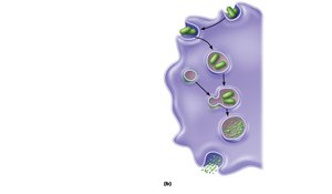
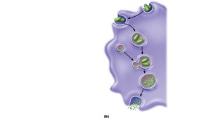
Phagocytosis is the process by which phagocytes engulf and destroy pathogens. The steps are:
Adherence: Phagocyte recognizes and adheres to pathogen's carbohydrate signature.
Engulfment: Phagocyte forms pseudopods to engulf the pathogen, creating a phagosome.
Fusion: Lysosome fuses with the phagosome, forming a phagolysosome.
Destruction: Toxic compounds and lysosomal enzymes destroy the pathogen.
Exocytosis: Indigestible material is expelled from the cell.
Clinical Example: Tuberculosis
Some bacteria, such as tuberculosis bacilli, resist digestion by macrophages and can remain alive inside, forming granulomas. If immunity decreases, these bacteria may become active and cause disease.
Helper T Cells and Respiratory Burst
Helper T cells can trigger macrophages to produce a respiratory burst, killing pathogens resistant to lysosomal enzymes by:
Releasing cell-killing free radicals
Producing oxidizing chemicals (e.g., H2O2)
Increasing pH and osmolarity of phagolysosome
Natural Killer (NK) Cells
Role and Mechanism
NK cells are nonphagocytic, large granular lymphocytes that police blood and lymph.
Kill cancer and virus-infected cells before the adaptive immune system is activated
Attack cells lacking "self" cell-surface receptors
Induce apoptosis in target cells
Secrete chemicals that enhance the inflammatory response
Inflammation: Tissue Response to Injury
Definition and Triggers
Inflammation is triggered whenever body tissues are injured by trauma, heat, chemicals, or infection.
Benefits of Inflammation
Prevents spread of damaging agents
Disposes of cell debris and pathogens
Alerts the adaptive immune system
Sets the stage for repair
Cardinal Signs of Acute Inflammation
Redness
Heat
Swelling
Pain
Impairment of function (sometimes)
Inflammatory Chemical Release
Chemicals such as histamine, kinins, prostaglandins, and complement are released by injured tissues, immune cells, or blood proteins.
Cause vasodilation of local arterioles
Make capillaries leaky
Attract leukocytes to the area
Trigger pain receptors and prompt release of more inflammatory chemicals
Vasodilation and Increased Vascular Permeability
Vasodilation: Causes hyperemia (increased blood flow), leading to redness and heat.
Increased Permeability: Causes exudate (fluid with clotting factors and antibodies) to leak into tissue, resulting in swelling (edema) and pain.
Benefits of Edema: Sweeps foreign material into lymphatic vessels, delivers clotting proteins and complement, forms fibrin mesh for repair, and isolates injured area.
Phagocyte Mobilization During Inflammation
Steps of Phagocyte Mobilization
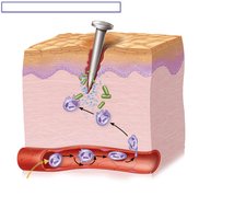
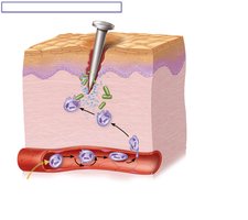
Leukocytosis: Neutrophils enter blood from bone marrow.
Margination: Neutrophils cling to capillary walls.
Diapedesis: Neutrophils flatten and squeeze out of capillaries.
Chemotaxis: Neutrophils follow chemical trail to site of injury.
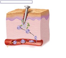
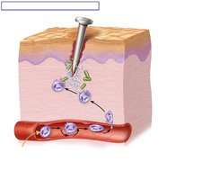
Summary Table: Inflammatory Response Steps
Step | Description |
|---|---|
Leukocytosis | Increase in white blood cells in bloodstream |
Margination | Leukocytes cling to capillary walls |
Diapedesis | Leukocytes pass through capillary walls |
Chemotaxis | Leukocytes follow chemical signals to site of injury |
Conclusion
The innate immune system provides rapid, nonspecific defense against pathogens through surface barriers, phagocytes, NK cells, and inflammation. Understanding these mechanisms is essential for assessing patient health and recognizing signs of a healthy immune response. Additional info: Academic context was added to clarify the steps of phagocytosis, the role of chemical barriers, and the clinical relevance of inflammation and phagocyte mobilization.
