 뒤로
Laboratory Study Guide: Endocrine Organs, Blood, and Blood Vessels
스터디 가이드 - 스마트 노트
 자료에 맞춘 맞춤형 노트, 핵심 정의, 예시, 맥락을 확장해 제공합니다.
Endocrine Organs
Overview of the Endocrine System
The endocrine system consists of glands and tissues that secrete hormones directly into the bloodstream to regulate various physiological processes. These hormones control growth, metabolism, reproduction, and homeostasis. - Primary endocrine organs: Hypothalamus, pituitary gland, thyroid gland, parathyroid glands, adrenal glands, pancreas, pineal gland, gonads (testis and ovary). - Secondary endocrine functions: Heart, thymus, digestive tract, adipose tissue, kidneys. - Functions: Regulation of blood pressure, immunity, digestion, metabolism, and reproductive processes. 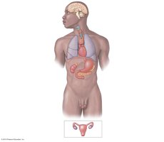
Hypothalamus and Pituitary Gland
The hypothalamus is a key regulatory center in the brain that controls the pituitary gland, which is often called the "master gland" due to its wide-ranging effects. - Hypothalamus: Produces antidiuretic hormone (ADH) and oxytocin, and releases regulatory hormones that control the anterior pituitary. - Pituitary gland: Divided into anterior and posterior lobes. The anterior lobe releases hormones affecting the thyroid, adrenal cortex, and reproductive organs. The posterior lobe releases ADH and oxytocin. - Adrenal medulla: Receives direct neural stimulation from the hypothalamus, releasing epinephrine and norepinephrine. 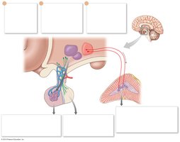
Thyroid and Parathyroid Glands
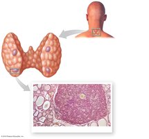
The thyroid gland is located in the neck and regulates metabolism, while the parathyroid glands control calcium balance. - Thyroid gland: Composed of follicles lined by simple cuboidal epithelium, containing colloid with thyroglobulin. C (clear) cells produce calcitonin. - Parathyroid glands: Small glands embedded in the posterior surface of the thyroid, secrete parathyroid hormone (PTH) to increase blood calcium levels. 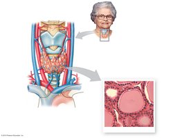
Pancreas and Pancreatic Islets
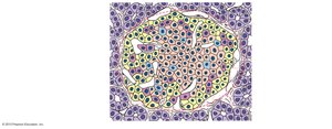
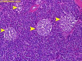
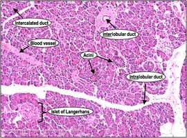
The pancreas has both endocrine and exocrine functions. Its endocrine portion, the pancreatic islets (Islets of Langerhans), regulates blood glucose. - Alpha cells: Produce glucagon, which increases blood glucose. - Beta cells: Produce insulin, which decreases blood glucose. - Pancreatic acini: Exocrine cells producing digestive enzymes. 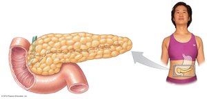
Pineal Gland
The pineal gland is located in the brain and secretes melatonin, which regulates circadian rhythms. - Melatonin: Hormone involved in sleep-wake cycles. 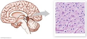
Adrenal Glands
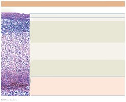
The adrenal glands are located above the kidneys and consist of two main regions: cortex and medulla. - Adrenal cortex: Produces mineralocorticoids (aldosterone), glucocorticoids (cortisol), and androgens. - Adrenal medulla: Produces epinephrine and norepinephrine. 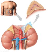
Blood
Components of Blood
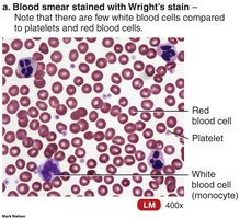
Blood is a connective tissue composed of plasma and formed elements. It transports nutrients, gases, wastes, and hormones throughout the body. - Plasma: The liquid portion, containing water, proteins (albumins, globulins, fibrinogen), electrolytes, nutrients, and wastes. - Formed elements: Red blood cells (RBCs), white blood cells (WBCs), and platelets. 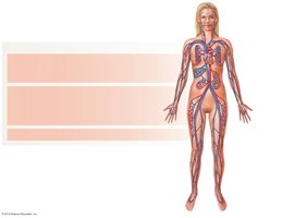
Red Blood Cells (Erythrocytes)
RBCs are specialized for oxygen transport due to their biconcave shape and hemoglobin content. - Shape: Biconcave disc increases surface area for gas exchange. - Function: Carry oxygen from lungs to tissues and carbon dioxide from tissues to lungs. 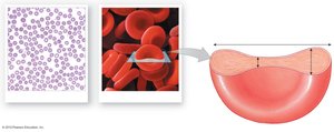
White Blood Cells (Leukocytes)
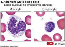
WBCs are involved in immune defense and are classified as granular or agranular. - Granular leukocytes: Neutrophils, eosinophils, basophils. - Agranular leukocytes: Monocytes, lymphocytes. - Functions: Phagocytosis, antibody production, inflammation, and defense against pathogens. 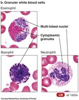
Blood Types and Antigens
Blood types are determined by the presence of surface antigens (A, B, AB, O) and antibodies in plasma. - Type A: Surface antigen A, anti-B antibodies. - Type B: Surface antigen B, anti-A antibodies. - Type AB: Both antigens, no antibodies. - Type O: No antigens, both antibodies. 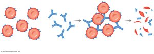
Cross-Reactions and Agglutination
When incompatible blood types are mixed, antibodies react with antigens, causing agglutination and hemolysis. - Agglutination: Clumping of RBCs due to antibody-antigen reaction. - Hemolysis: Destruction of RBCs following agglutination. 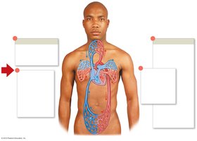
Blood Vessels
Structure of Blood Vessels
Blood vessels are classified as arteries, veins, and capillaries, each with distinct structural features. - Arteries: Thick walls, high pressure, carry blood away from the heart. - Veins: Thinner walls, lower pressure, carry blood toward the heart, often contain valves. - Capillaries: Thin walls for exchange of gases, nutrients, and wastes. 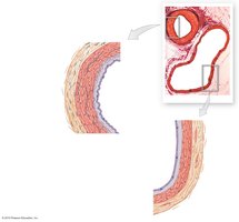
Layers of Blood Vessels
Blood vessels have three main layers: tunica intima, tunica media, and tunica externa. - Tunica intima: Innermost layer, endothelium. - Tunica media: Middle layer, smooth muscle and elastic fibers. - Tunica externa: Outermost layer, connective tissue. 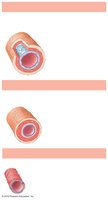
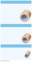
Venous Valves and Blood Flow
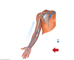
Veins contain valves to prevent backflow and ensure unidirectional blood flow toward the heart. - Valves: Open above contracting muscle, closed below to prevent backflow. 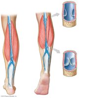
Capillary Beds and Blood Flow Regulation
Capillary beds are networks of capillaries that regulate blood flow via precapillary sphincters and arteriovenous anastomoses. - Precapillary sphincters: Control blood flow into capillaries. - Arteriovenous anastomosis: Direct connection between arterioles and venules. 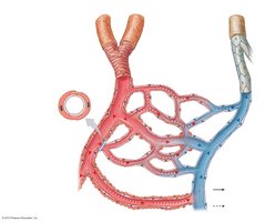
Circuits of the Cardiovascular System
The cardiovascular system is divided into pulmonary and systemic circuits. - Pulmonary circuit: Carries blood from the heart to the lungs and back for gas exchange. - Systemic circuit: Carries blood from the heart to the rest of the body and back. 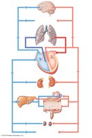
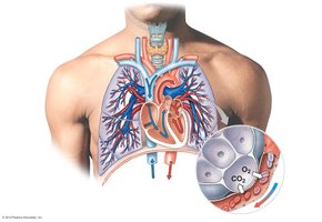
Major Arteries and Veins
The body contains numerous arteries and veins, each serving specific regions. - Arteries: Aorta, carotid, subclavian, axillary, brachial, radial, ulnar, femoral, popliteal, tibial, etc. - Veins: Jugular, subclavian, cephalic, basilic, femoral, saphenous, popliteal, etc. 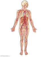
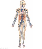
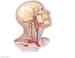
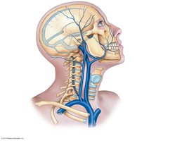
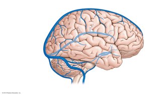
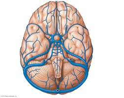
Cerebral Circulation
The brain receives blood through the cerebral arterial circle (Circle of Willis) and is drained by dural sinuses. - Circle of Willis: Provides collateral circulation to the brain. - Dural sinuses: Drain venous blood from the brain. 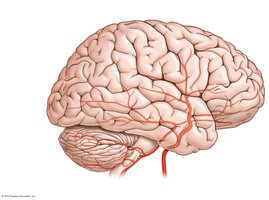
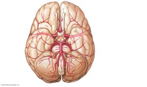
Abdominal Circulation
The abdominal aorta and its branches supply blood to abdominal organs, while the hepatic portal system drains blood from the digestive tract to the liver. - Major arteries: Celiac trunk, superior mesenteric, inferior mesenteric, renal, gonadal, common iliac. - Hepatic portal system: Blood from digestive organs passes through the liver for processing. 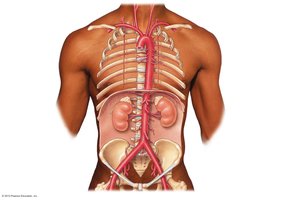
Arteries and Veins of the Lower Limb
The lower limb is supplied by the femoral, popliteal, tibial, and fibular arteries and drained by corresponding veins. - Arteries: Femoral, popliteal, anterior tibial, posterior tibial, fibular, dorsalis pedis, plantar arch. - Veins: Femoral, great saphenous, small saphenous, popliteal, anterior tibial, posterior tibial, fibular, dorsal and plantar venous arches.
Additional info:
Some histological images and diagrams were interpreted based on standard anatomical and physiological knowledge to provide context and clarity for study purposes.
