 뒤로
Membrane Transport & Excitable Cells: Study Guide for Anatomy & Physiology
스터디 가이드 - 스마트 노트
 자료에 맞춘 맞춤형 노트, 핵심 정의, 예시, 맥락을 확장해 제공합니다.
Membrane Structure and Function
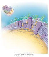
Fluid Mosaic Model of the Plasma Membrane
The plasma membrane is a dynamic structure essential for cellular integrity and function. It is described by the fluid mosaic model, which highlights its composition and flexibility.
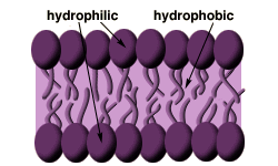
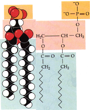
Phospholipid Bilayer: The fundamental structure consists of two layers of phospholipids. Each phospholipid has a hydrophilic (water-loving) head and hydrophobic (water-fearing) tails.
Hydrophilic vs. Hydrophobic: Hydrophilic heads face outward toward aqueous environments, while hydrophobic tails face inward, away from water.
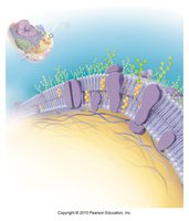
Integral Proteins: Span the membrane, possessing both hydrophilic and hydrophobic regions. They function as channels, carriers, or receptors.
Peripheral Proteins: Attach to integral proteins, often on the internal face, and serve as enzymes, attachment points, or receptors.
Cytoskeleton: Anchors to the plasma membrane, providing structural support.
Glycocalyx: A carbohydrate-rich layer on the membrane's exterior, important for cell recognition and immune response.
Cholesterol: Stabilizes membrane structure and modulates fluidity.
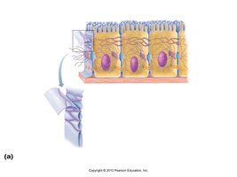
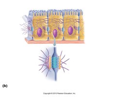
Membrane Junctions
Specialized junctions between cells allow for communication, adhesion, and barrier formation.
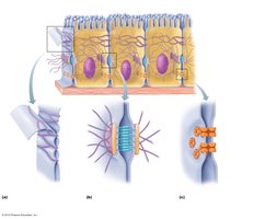
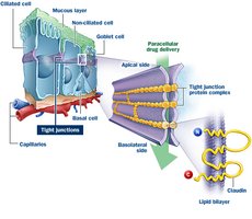
Tight Junctions: Fusion of adjacent membranes prevents passage of molecules between cells, crucial in tissues like the digestive tract.
Desmosomes: Anchoring junctions that resist mechanical stress, found in tissues subject to stretching (e.g., skin, heart).
Gap Junctions: Channels that allow passage of ions and small molecules, important in electrically excitable tissues.
Functions of Plasma Membrane Proteins
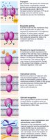
Membrane proteins are vital for cellular communication, transport, and structural integrity.
Transport: Facilitate movement of substances across the membrane.
Enzymatic Activity: Catalyze reactions at the membrane surface.
Signal Transduction: Act as receptors for hormones and other signals.
Intercellular Joining: Form junctions between cells.
Cell-Cell Recognition: Allow cells to identify each other.
Attachment to ECM: Anchor cells to the extracellular matrix.
Membrane Transport Mechanisms
Passive Transport
Passive transport does not require energy and relies on concentration gradients.
Diffusion: Movement of molecules from high to low concentration due to kinetic energy.
Simple Diffusion: Lipid-soluble molecules (O2, CO2, fats, urea, alcohol) pass directly through the membrane.
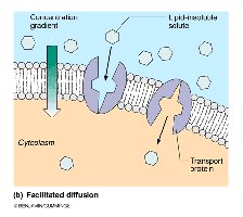
Facilitated Diffusion: Water-soluble substances use specific carrier or channel proteins. Movement is down the concentration gradient and does not require ATP.
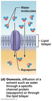
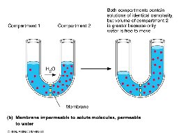
Osmosis: Diffusion of water across a semipermeable membrane, often through aquaporins.
Osmolarity and Tonicity
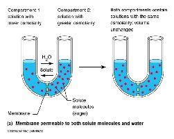
Osmolarity: Total concentration of solute particles in a solution, measured in mOsmol/L.
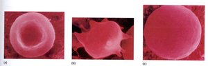
Tonicity: The effect of a solution on cell shape by altering water content, determined by nonpenetrating solutes.
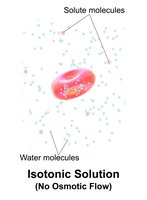
Isotonic: No net water movement; cell shape remains unchanged.
Hypertonic: Water leaves the cell; cell shrinks.
Hypotonic: Water enters the cell; cell swells and may lyse.
Active Transport
Active transport requires ATP to move substances against their concentration gradients or when substances are too large or lipid-insoluble.
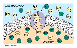
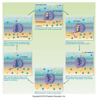
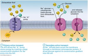
Primary Active Transport: Direct use of ATP, e.g., Na+/K+ ATPase pump.
Secondary Active Transport: Uses gradients established by primary active transport to drive cotransport (symport or antiport).
Vesicular Transport
Vesicular transport involves the movement of large particles or volumes via vesicles and requires ATP.
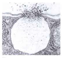
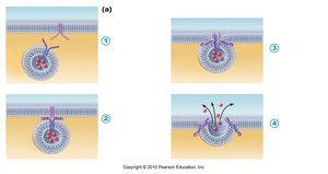
Exocytosis: Vesicles fuse with the plasma membrane to release contents outside the cell (e.g., secretion of hormones, neurotransmitters).
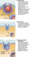
Endocytosis: Vesicles form to bring substances into the cell. Includes phagocytosis (solid particles), pinocytosis (fluid), and receptor-mediated endocytosis (specific macromolecules).
Excitable Cells: Neurons
Structure and Function of Neurons
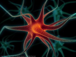
Neurons are specialized cells for electrical impulse conduction. They possess unique features and regions for reception, propagation, and transmission.
Extreme Longevity: Neurons can last a lifetime if properly nourished.
Amitotic: Most neurons do not divide after development.
High Metabolic Rate: Require continuous supply of O2 and glucose.
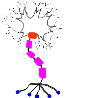
Cell Body: Contains nucleus, biosynthetic center, and organelles.
Dendrites: Receptive region; conduct impulses toward cell body.
Axon: Conducting region; transmits impulses away from cell body.
Axon Terminals: Secretory region; release neurotransmitters.

Neuron Processes and Transport
Tract: Bundle of nerve processes in the CNS.
Nerve: Bundle of nerve processes in the PNS.
Anterograde Transport: Moves materials from cell body to axon terminals.
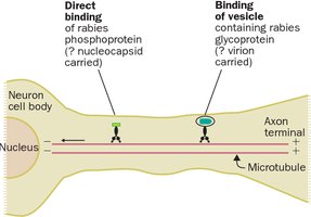
Retrograde Transport: Moves materials from axon terminals to cell body; used by viruses and toxins.
Clinical Note
Certain viruses (e.g., polio, rabies, herpes simplex) and toxins (e.g., tetanus) exploit retrograde transport to reach neuron cell bodies, leading to neurological disease.
Summary Table: Types of Membrane Transport
Type | Energy Required | Direction | Example |
|---|---|---|---|
Simple Diffusion | No | Down gradient | O2, CO2 |
Facilitated Diffusion | No | Down gradient | Glucose, ions |
Osmosis | No | Down gradient | Water |
Primary Active Transport | Yes (ATP) | Against gradient | Na+/K+ pump |
Secondary Active Transport | Indirect (ATP) | Against gradient | Na+-glucose cotransport |
Vesicular Transport | Yes (ATP) | Bulk movement | Exocytosis, endocytosis |
Key Equations
Osmolarity Calculation
Osmolarity is the sum of all solute particles in solution.
Example: 1 mM NaCl = 2 mOsm (Na+ and Cl- dissociate)
1 mM CaCl2 = 3 mOsm (Ca2+ and 2 Cl-)
Na+/K+ ATPase Pump
For every cycle: 3 Na+ out, 2 K+ in, 1 ATP hydrolyzed.
Additional info:
Membrane transport is fundamental for maintaining cellular homeostasis, electrical excitability, and communication.
Neurons rely on these mechanisms for rapid signaling and integration in the nervous system.
