 뒤로
Muscle Structure, Function, and Physiology Study Guide
스터디 가이드 - 스마트 노트
 자료에 맞춘 맞춤형 노트, 핵심 정의, 예시, 맥락을 확장해 제공합니다.
Q1. Which of the following cells are excitable?
Background
Topic: Excitability of Cells
This question tests your understanding of which cell types can respond to stimuli by generating action potentials.
Key Terms:
Excitable cells: Cells that can generate and propagate electrical signals (action potentials).
Examples: Cardiac muscle cells, skeletal muscle cells, smooth muscle cells, neurons.
Step-by-Step Guidance
Recall the definition of excitability in physiology: the ability to respond to a stimulus and generate an action potential.
Think about the main cell types involved in muscle contraction and nervous system signaling.
Review which cells are responsible for voluntary and involuntary movements, and which transmit electrical signals.
Consider whether all listed cell types share this property or if only some do.
Try solving on your own before revealing the answer!
Final Answer: All of the above
Cardiac, skeletal, and smooth muscle cells, as well as neurons, are all excitable because they can generate action potentials in response to stimuli.
Q2. The pulling effort of a muscle is transferred via a ______ from the muscle to the bone.
Background
Topic: Muscle Attachments
This question is about how muscles connect to bones and transmit force to produce movement.
Key Terms:
Tendon: Dense connective tissue that attaches muscle to bone.
Ligament: Connects bone to bone.
Belly: The thick, central part of a muscle.
Step-by-Step Guidance
Recall the structure of a skeletal muscle and its attachments.
Identify which structure is responsible for transmitting force from muscle contraction to bone movement.
Compare the roles of tendons, ligaments, and muscle belly in movement.
Try solving on your own before revealing the answer!
Final Answer: Tendon
The tendon is the structure that transfers the pulling effort of a muscle to the bone, enabling movement.
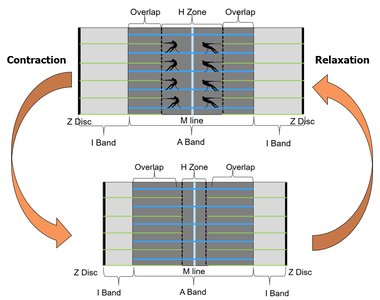
Q3. The _____ contains only actin, while the _____ contains only myosin. These regions get _____ during contraction.
Background
Topic: Sarcomere Structure
This question tests your knowledge of the arrangement of actin and myosin filaments within the sarcomere and how these regions change during contraction.
Key Terms:
I band: Region with only actin (thin filaments).
H zone: Region with only myosin (thick filaments).
Contraction: The process where sarcomere shortens, changing the size of these regions.
Step-by-Step Guidance
Review the structure of the sarcomere, focusing on the I band and H zone.
Recall which filaments are present in each region.
Think about what happens to these regions when the muscle contracts.
Set up your answer by matching the regions to their filament content and describing the change during contraction.
Try solving on your own before revealing the answer!
Final Answer: I band; H zone; smaller
The I band contains only actin, the H zone contains only myosin, and both regions get smaller during contraction as the filaments overlap more.
Q4. During contraction, the length of the sarcomere _____ and the length of the myofilaments _____.
Background
Topic: Muscle Contraction Mechanics
This question is about the changes in sarcomere and myofilament length during muscle contraction.
Key Terms:
Sarcomere: The functional unit of muscle contraction.
Myofilaments: Actin and myosin filaments within the sarcomere.
Step-by-Step Guidance
Recall the sliding filament theory: actin and myosin slide past each other, causing the sarcomere to change length.
Think about whether the filaments themselves change length or just their overlap.
Set up your answer by describing the change in sarcomere length and myofilament length during contraction.
Try solving on your own before revealing the answer!
Final Answer: decreases; stays the same
During contraction, the sarcomere shortens (decreases in length), but the myofilaments themselves do not change length; they slide past each other.
Q5. During relaxation, the zone of overlap ______ and the Z discs move ______.
Background
Topic: Muscle Relaxation
This question tests your understanding of what happens to the sarcomere structure during muscle relaxation.
Key Terms:
Zone of overlap: Area where actin and myosin filaments overlap.
Z discs: Boundaries of the sarcomere.
Step-by-Step Guidance
Recall what happens to the sarcomere when the muscle relaxes.
Think about the relationship between the zone of overlap and the Z discs during relaxation.
Set up your answer by describing the changes in these regions.
Try solving on your own before revealing the answer!
Final Answer: decreases; farther apart
During relaxation, the zone of overlap decreases as the filaments slide apart, and the Z discs move farther apart as the sarcomere lengthens.
Q6. Which of the following is mismatched?
Background
Topic: Muscle Physiology Phases
This question tests your understanding of the phases of muscle contraction: excitation, contraction, and relaxation.
Key Terms:
Excitation: Initiation of action potential and neurotransmitter release.
Contraction: Cross-bridge cycling and sarcomere shortening.
Relaxation: Sequestering calcium and muscle lengthening.
Step-by-Step Guidance
Review the definitions and main events of each phase.
Compare the descriptions given in the question to the actual events of each phase.
Identify which pairing does not accurately match the phase to its event.
Try solving on your own before revealing the answer!
Final Answer: Excitation: Generation of cross bridges
Excitation does not involve the generation of cross bridges; this occurs during contraction. Excitation is about action potential and neurotransmitter release.
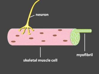
Q7. The axon contains vesicles of _____ and the _____ contains receptors for this neurotransmitter.
Background
Topic: Neuromuscular Junction
This question tests your knowledge of neurotransmitter release and receptor location at the neuromuscular junction.
Key Terms:
Acetylcholine (Ach): Neurotransmitter released by motor neurons.
Motor end plate: Region of muscle cell membrane with Ach receptors.
Step-by-Step Guidance
Recall the process of neurotransmitter release at the neuromuscular junction.
Identify where Ach is stored and where its receptors are located.
Set up your answer by matching the neurotransmitter to its storage site and receptor location.
Try solving on your own before revealing the answer!
Final Answer: Ach; motor end plate
The axon contains vesicles of acetylcholine, and the motor end plate contains receptors for acetylcholine.
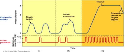
Q8. Which of the following would most likely lead to fatigue due to continuous contraction (no relaxation) in the muscle fiber?
Background
Topic: Muscle Fatigue and Tension
This question tests your understanding of muscle contraction types and their relationship to fatigue.
Key Terms:
Wave summation: Increased tension due to repeated stimulation.
Incomplete tetanus: Sustained contraction with some relaxation.
Tetanus: Continuous, maximal contraction with no relaxation.
Step-by-Step Guidance
Review the definitions of wave summation, incomplete tetanus, and tetanus.
Consider which type results in the greatest and most sustained tension.
Think about which condition would most likely cause fatigue due to lack of relaxation.
Try solving on your own before revealing the answer!
Final Answer: Tetanus
Tetanus is a state of continuous contraction with no relaxation, which most likely leads to fatigue in the muscle fiber.
Q9. You are picking up a heavy object. Which of the following motor units would you want to recruit in the skeletal muscles of your forearm?
Background
Topic: Motor Unit Recruitment
This question tests your understanding of how motor units are recruited for different tasks.
Key Terms:
Motor unit: A motor neuron and all the muscle fibers it innervates.
Recruitment: Activation of additional motor units to increase muscle force.
Step-by-Step Guidance
Recall the concept of motor unit recruitment and its role in generating force.
Think about the difference between small and large motor units.
Consider which type of motor unit would be most effective for lifting a heavy object.

Try solving on your own before revealing the answer!
Final Answer: Larger motor units that have one nerve and many muscle fibers
Recruiting larger motor units allows for greater force generation, which is necessary for lifting heavy objects.
