 뒤로
뒤로Muscle Tissue and Physiology: Structure, Function, and Contraction
스터디 가이드 - 스마트 노트
 자료에 맞춘 맞춤형 노트, 핵심 정의, 예시, 맥락을 확장해 제공합니다.
Muscle Tissue and Physiology
Introduction to Muscles and Muscle Tissue
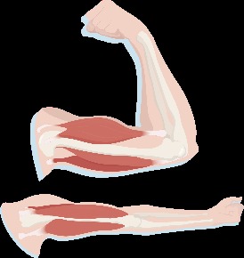
Muscle tissue is specialized for contraction, enabling movement by converting chemical energy into mechanical energy. Muscles generate force and are essential for locomotion, posture, and vital functions such as heartbeats and digestion.
Contractility: The ability of muscle tissue to forcibly shorten, producing movement.
Extensibility: The ability to stretch without being damaged.
Elasticity: The ability to return to original shape after being stretched.
Excitability: The ability to respond to stimuli, usually from nerves.
Types of Muscle Tissue
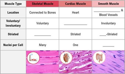
There are three types of muscle tissue in the human body: skeletal, cardiac, and smooth. Each type has distinct structural and functional characteristics.
Skeletal Muscle: Voluntary, striated, attached to bones, multinucleated.
Cardiac Muscle: Involuntary, striated, found in the heart, single nucleus per cell.
Smooth Muscle: Involuntary, non-striated, found in walls of hollow organs and blood vessels, single nucleus per cell.
Muscle Type | Location | Voluntary/Involuntary | Striated | Nuclei per Cell |
|---|---|---|---|---|
Skeletal Muscle | Connected to Bones | Voluntary | Striated | Many |
Cardiac Muscle | Heart | Involuntary | Striated | One |
Smooth Muscle | Blood Vessels & Organs | Involuntary | Non-Striated | One |
Structure of a Skeletal Muscle
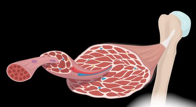
Skeletal muscles are organized into hierarchical structures, including muscle fibers, fascicles, and connective tissue layers. This organization allows for efficient force generation and transmission.
Muscle Fiber: A single, long, multinucleated cell surrounded by endomysium.
Fascicle: Bundle of muscle fibers surrounded by perimysium.
Muscle: Bundle of fascicles surrounded by epimysium.
Connective Tissue: Tendons (cord-like) and aponeuroses (sheet-like) connect muscles to bones.
The Muscle Fiber: Cellular Structure
Muscle fibers contain specialized organelles and structures for contraction. The plasma membrane (sarcolemma) wraps the fiber, and T-tubules allow signals to reach deep into the cell. Myofibrils are rod-shaped organelles containing myofilaments (actin and myosin).
Sarcolemma: Plasma membrane of the muscle fiber.
T-Tubules: Invaginations of the sarcolemma that transmit action potentials.
Sarcoplasmic Reticulum: Stores and releases calcium ions, surrounding myofibrils.
Myofibrils: Contain contractile proteins (actin and myosin).
Sliding Filament Theory and the Sarcomere
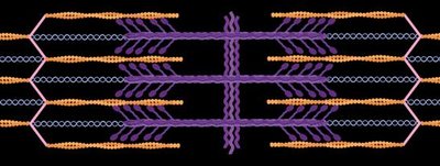
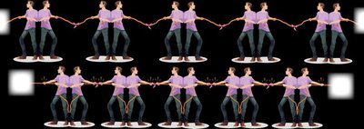
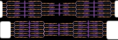
The sliding filament theory explains how muscles contract. The sarcomere is the basic contractile unit, composed of thick (myosin) and thin (actin) filaments. During contraction, myosin pulls actin toward the center, shortening the sarcomere.
Myosin: Thick filament, anchored to the center (M line) of the sarcomere.
Actin: Thin filament, anchored to the ends (Z disc) of the sarcomere.
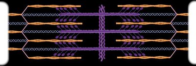
Contraction: Filaments slide past each other; overlap increases, but filament length remains constant.
Proteins of the Sarcomere
The sarcomere contains contractile, regulatory, and structural proteins. These proteins coordinate contraction and maintain structural integrity.
Contractile Proteins:
Myosin: Thick filament, many-headed structure.
Actin: Thin filament.
Regulatory Proteins:
Tropomyosin: Wraps actin, blocks myosin binding sites.
Troponin: Binds calcium, moves tropomyosin to expose binding sites.
Structural Proteins:
Titin: Elastic filament, helps sarcomere retain shape.
Structure of the Sarcomere: Bands, Zones, Discs & Lines
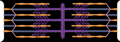
The sarcomere is divided into regions based on appearance under a microscope. These regions change during contraction.
Z Disc: End of the sarcomere, anchors actin.
M Line: Center, anchors myosin.
I Band: Area with only actin.
A Band: Area with both actin and myosin.
H Zone: Center region with only myosin.
Region | Description |
|---|---|
A Band | Contains both actin and myosin; does not change length during contraction. |
I Band | Contains only actin; shortens during contraction. |
H Zone | Contains only myosin; shortens during contraction. |
Z Disc | Anchors actin; moves closer together during contraction. |
M Line | Anchors myosin; remains central. |
Steps of Muscle Contraction
Muscle contraction involves a series of steps, including signal transmission, excitation-contraction coupling, and the cross-bridge cycle.
Events at the Neuromuscular Junction
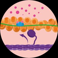
Neurotransmitter Release: Acetylcholine is released from the motor neuron, crosses the synaptic cleft, and binds to receptors on the sarcolemma.
Action Potential: Initiated in the muscle fiber, propagates along the sarcolemma and T-tubules.
Calcium Release: Sarcoplasmic reticulum releases Ca2+ ions.
Excitation-Contraction Coupling
Action Potential: Travels down the sarcolemma and T-tubules.
Calcium Binding: Ca2+ binds to troponin, moving tropomyosin and exposing myosin binding sites on actin.
Cross-Bridge Formation: Myosin binds to actin, initiating contraction.
Cross Bridge Cycle
Myosin Head Binds to Actin: Forms a cross-bridge.
Power Stroke: Myosin pulls actin, shortening the sarcomere.
ATP Binding: Myosin releases actin when ATP binds.
ATP Hydrolysis: Myosin head returns to cocked position, ready for another cycle.
Key Equation:
Neurotransmitters & Action Potentials
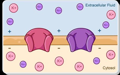
Neurotransmitters such as acetylcholine are essential for signal transmission at the neuromuscular junction. Action potentials are changes in membrane polarization that propagate along the muscle fiber.
Polarization: Resting state, inside of cell is negatively charged.
Depolarization: Na+ enters cell, inside becomes more positive.
Repolarization: K+ exits cell, charge is restored.
Summary Table: Sarcomere Regions and Changes During Contraction
Region | Change During Contraction |
|---|---|
A Band | No change |
I Band | Shortens |
H Zone | Shortens |
Z Disc | Moves closer together |
M Line | Remains central |
Summary Table: Muscle Contraction Steps
Step | Description |
|---|---|
1 | Action potential arrives at axon terminal |
2 | Voltage-gated Ca2+ channels open |
3 | Ca2+ enters, acetylcholine released |
4 | ACh binds to sarcolemma receptors |
5 | Na+ channels open, action potential starts in muscle fiber |
6 | ACh broken down, signal ends |
Key Terms
Sarcomere: Basic contractile unit of muscle fiber.
Myofibril: Rod-shaped organelle containing myofilaments.
Actin: Thin filament protein.
Myosin: Thick filament protein.
Tropomyosin: Regulatory protein blocking myosin binding sites.
Troponin: Regulatory protein binding calcium.
Titin: Structural protein providing elasticity.
Sarcolemma: Muscle fiber plasma membrane.
Sarcoplasmic Reticulum: Stores and releases calcium ions.
Neuromuscular Junction: Synapse between motor neuron and muscle fiber.
Acetylcholine: Neurotransmitter at neuromuscular junction.
Additional info: Academic context was added to clarify the structure and function of muscle tissue, the sliding filament theory, and the steps of muscle contraction. Tables were reconstructed for comparison and process steps. Images were included only when directly relevant to the explanation.
