 뒤로
뒤로Muscle Tissue and Physiology: Study Notes for Anatomy & Physiology
스터디 가이드 - 스마트 노트
 자료에 맞춘 맞춤형 노트, 핵심 정의, 예시, 맥락을 확장해 제공합니다.
Muscle Tissue: Properties and Overview
Properties of Muscle Cells
Muscle cells possess unique properties that enable them to perform their functions in the body. These properties are essential for movement, stability, and other physiological processes.
Contractility: The ability of muscle cells to shorten forcefully when stimulated.
Excitability: The capacity to respond to stimuli, usually from the nervous system.
Conductivity: The ability to transmit electrical signals along the cell membrane.
Distensibility: The ability to stretch without being damaged.
Elasticity: The ability to return to original shape after being stretched or contracted.
Structure and Function of Skeletal Muscle Fibers
Sliding-Filament Theory
The sliding-filament theory explains how muscles contract at the molecular level. It involves the interaction between actin and myosin filaments within the sarcomere, the functional unit of muscle fibers.
Actin: Thin filament that provides sites for myosin attachment.
Myosin: Thick filament with heads that bind to actin and pull it during contraction.
ATP: Provides energy for myosin head movement and detachment.
Electrically Excitable Cells
Membrane Potentials
Muscle cells maintain a resting membrane potential, which is essential for their excitability and function.
Resting Potential: The polarized state of the cell membrane, typically around -70 mV.
Channels and Gradients:
Leak Channels: Allow passive movement of ions.
Gated Channels: Open in response to specific stimuli.
Electrochemical Gradient: Combination of concentration and electrical gradients.
Action Potentials:
Resting Stage: Cell is polarized.
Depolarization Stage: Sodium ions enter, making the inside more positive.
Repolarization Stage: Potassium ions exit, restoring negative charge.
Propagation: Action potential travels along the membrane.
Process of Skeletal Muscle Contraction and Relaxation
The Neuromuscular Junction (NMJ)
The NMJ is the site where a motor neuron communicates with a muscle fiber to initiate contraction.
Motor Neuron: Transmits the signal to the muscle.
Anatomy:
Axon Terminal: Releases neurotransmitter.
Synaptic Cleft: Space between neuron and muscle.
Motor End Plate: Region of muscle membrane with receptors.
Skeletal Muscle Contraction
Excitation Phase:
Action potential arrives at axon terminal, opening calcium channels.
Calcium triggers exocytosis of synaptic vesicles.
Acetylcholine (ACh) released into synaptic cleft.
ACh binds to receptors on motor end plate.
Sodium channels open, sodium enters muscle fiber.
Depolarization creates end-plate potential.
Excitation-Contraction Coupling:
End-plate potential stimulates action potential.
Action potential propagates down T-tubules.
Calcium channels in sarcoplasmic reticulum open, calcium enters cytosol.
Calcium binds to troponin.
Tropomyosin moves, exposing actin sites.
Contraction Phase:
ATP hydrolysis cocks myosin head.
Myosin head binds to actin.
Power stroke: Myosin pulls actin toward sarcomere center.
ATP breaks myosin-actin attachment.
Muscle Relaxation:
Acetylcholinesterase degrades ACh, repolarization occurs.
Sarcolemma returns to resting potential, calcium channels close.
Calcium pumped back into SR.
Troponin shifts, tropomyosin blocks actin sites, muscle relaxes.
Relaxation is passive.
Energy Sources for Muscle Contraction
Immediate Energy
Creatine Phosphate: Provides rapid ATP regeneration for short bursts of activity.
Anaerobic Catabolism (Glycolysis)
Glucose Sources: Blood glucose and glycogen.
Anaerobic: Does not require oxygen.
Fate of Pyruvate: Converted to lactic acid if oxygen is absent.
Aerobic Catabolism
Fuel Sources: Glucose, fatty acids, amino acids.
Aerobic: Requires oxygen; myoglobin stores oxygen in muscle.
Long Lasting: Supports prolonged activity.
Muscle Tension at the Fiber Level
Muscle Twitch
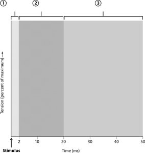
A muscle twitch is a single contraction cycle in response to a stimulus. It consists of three phases:
Latent Period: Time between stimulus and contraction onset.
Contraction Period: Muscle generates tension.
Relaxation Period: Muscle returns to resting state.
Refractory Period: Time during which muscle cannot respond to another stimulus.
Tension Production and Summation
Wave Summation: Increased tension due to repeated stimulation.
Unfused (Incomplete) Tetanus: Partial relaxation between stimuli.
Fused (Complete) Tetanus: No relaxation, sustained contraction.
Length-Tension Relationship
The amount of tension a muscle fiber can produce depends on its length at the time of stimulation.
Classes of Skeletal Muscle Fibers
Type I Fibers:
Slow twitch
Oxidative fibers
Fatigue resistant
Type II Fibers:
Fast twitch
Glycolytic fibers
Fatigue quickly
Muscle Tension at the Organ Level
Motor Units
A motor unit consists of a motor neuron and all the muscle fibers it innervates. Recruitment of motor units increases muscle tension.
Muscle Tone:
Hypotonia: Reduced muscle tone.
Hypertonia: Increased muscle tone.
Types of Contractions:
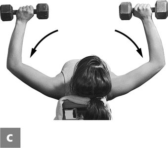
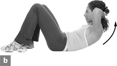
Isotonic: Muscle changes length.
Concentric: Muscle shortens.
Eccentric: Muscle lengthens.
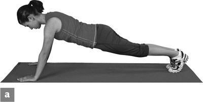
Isometric: Muscle length does not change.
Muscle Performance
Physical Training
Endurance Training: Increases oxidative capacity and fatigue resistance.
Resistance Training: Increases muscle strength and size.
Disuse: Leads to muscle atrophy and decreased performance.
Muscle Fatigue
Causes:
Depletion of energy sources
Accumulation of metabolic byproducts
Impaired calcium handling
Recovery Period
Increased Ventilation: After exercise, increased breathing helps restore oxygen levels and remove excess carbon dioxide.
Smooth and Cardiac Muscle Compared to Skeletal Muscle
Smooth Muscle
Similarities: Contractile proteins, excitability, and ability to generate tension.
Differences: Non-striated, involuntary, slower contraction, found in walls of hollow organs.
Cardiac Muscle
Similarities: Striated, involuntary, uses sliding-filament mechanism.
Differences: Branched cells, intercalated discs, found only in the heart, rhythmic contractions.
Key Equations
ATP Hydrolysis
ATP is hydrolyzed to provide energy for muscle contraction:
Creatine Phosphate Reaction
Creatine phosphate regenerates ATP:
Muscle Twitch Phases Table
Phase | Description |
|---|---|
Latent Period | Time between stimulus and contraction onset |
Contraction Period | Muscle generates tension |
Relaxation Period | Muscle returns to resting state |
Refractory Period | Muscle cannot respond to another stimulus |
Muscle Fiber Types Table
Type | Speed | Metabolism | Fatigue Resistance |
|---|---|---|---|
Type I | Slow | Oxidative | High |
Type II | Fast | Glycolytic | Low |
Types of Muscle Contractions Table
Type | Length Change | Example |
|---|---|---|
Isotonic Concentric | Shortens | Raising weights, sit-up |
Isotonic Eccentric | Lengthens | Lowering weights |
Isometric | No change | Plank position |
Additional info: Academic context and explanations were expanded for clarity and completeness. Images were included only where directly relevant to muscle twitch phases and types of muscle contractions.
