 뒤로
Muscle Tissue: Structure and Function in Anatomy & Physiology
스터디 가이드 - 스마트 노트
 자료에 맞춘 맞춤형 노트, 핵심 정의, 예시, 맥락을 확장해 제공합니다.
Muscle Tissue
Types of Muscle Tissue
Muscle tissue is a primary tissue in the human body, specialized for contraction and movement. There are three main types of muscle tissue:
Skeletal muscle: Responsible for voluntary movements by pulling on bones.
Cardiac muscle: Found only in the heart, controls rhythmic contractions.
Smooth muscle: Controls involuntary movements within internal organs.
Functions of Muscle Tissue
Muscle tissue performs several essential functions:
Producing movement: Skeletal muscles move the body.
Maintaining posture: Stabilizes body position.
Supporting soft tissues: Protects internal organs.
Guarding entrances and exits: Controls passage through body openings.
Maintaining body temperature: Generates heat during contraction.
Storing nutrients: Stores glycogen and other nutrients.
Organization of Skeletal Muscle
Structural Hierarchy
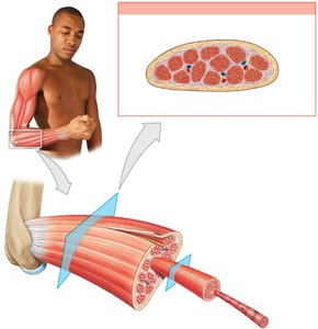
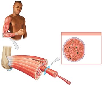
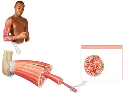
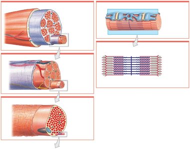
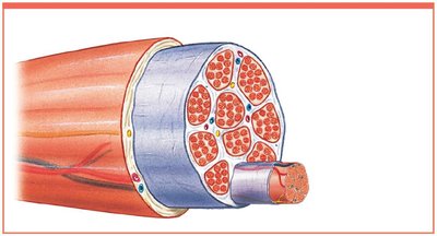
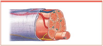
Skeletal muscle is organized into several structural levels, each surrounded by connective tissue:
Epimysium: Surrounds the entire muscle.
Perimysium: Surrounds bundles of muscle fibers called fascicles.
Endomysium: Surrounds individual muscle fibers.
Connective Tissue and Attachments
Collagen fibers from all three layers merge to form tendons (bundles) or aponeuroses (sheets), which attach muscles to bones.
Skeletal Muscle Fibers
Development and Structure
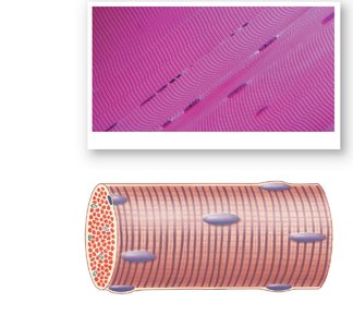
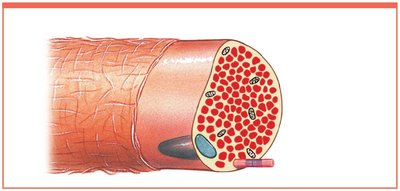
Skeletal muscle fibers are large, multinucleate cells formed by the fusion of embryonic cells called myoblasts.
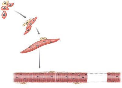
Each fiber contains hundreds of nuclei.
Striated appearance due to organized arrangement of contractile proteins.
Key Components
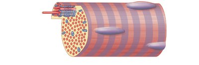
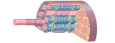
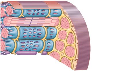
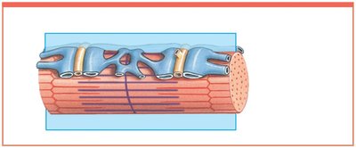
Sarcolemma: Plasma membrane of the muscle fiber.
Sarcoplasm: Cytoplasm of the muscle fiber.
Transverse tubules (T tubules): Invaginations of the sarcolemma that transmit action potentials.
Sarcoplasmic reticulum (SR): Specialized for storage and release of calcium ions.
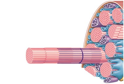
Myofibrils: Bundles of protein filaments responsible for contraction.
Sarcomere Structure
Functional Unit of Muscle
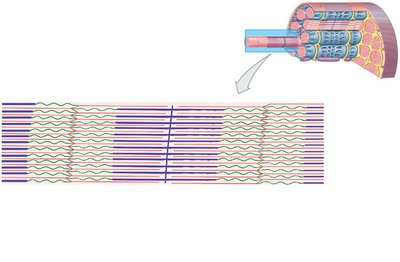
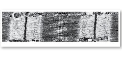
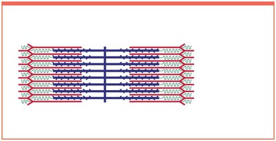
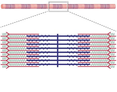
The sarcomere is the smallest functional unit of a muscle fiber. Its arrangement of thick and thin filaments produces the striated pattern and enables contraction.
A band: Dark region containing thick filaments.
I band: Light region containing thin filaments.
H band: Area with only thick filaments.
M line: Center of the A band, stabilizes thick filaments.
Z line: Boundary between sarcomeres, anchors thin filaments.
Titin: Elastic protein maintaining alignment and restoring length.
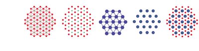
Levels of Functional Organization
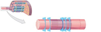
Muscle structure is organized from the whole muscle down to the sarcomere:
Skeletal muscle → Muscle fascicle → Muscle fiber → Myofibril → Sarcomere
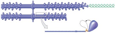
Myofilaments: Thin and Thick Filaments
Thin Filaments
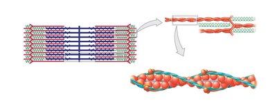
Composed of F-actin (filamentous actin), nebulin, tropomyosin, and troponin.
Active sites on G-actin bind to myosin heads.
Tropomyosin covers active sites, preventing interaction.
Troponin binds tropomyosin, G-actin, and Ca2+.
Thick Filaments
Composed of myosin molecules (each with a tail and two heads).
Myosin heads project toward thin filaments and form cross-bridges during contraction.
Core of titin provides elasticity.
Sliding-Filament Theory
Mechanism of Contraction
Muscle contraction occurs as thin filaments slide toward the center of the sarcomere, causing:
H and I bands to narrow
Zones of overlap to widen
Z lines to move closer together
A band width remains constant
Neuromuscular Junction and Excitation-Contraction Coupling
Neuromuscular Junction (NMJ)
The NMJ is the synapse between a motor neuron and a skeletal muscle fiber. The process involves:
Release of acetylcholine (ACh) from the neuron
ACh binds to receptors on the muscle fiber, opening Na+ channels
Depolarization generates an action potential
Excitation-Contraction Coupling
Action potential travels along T tubules to the triads
Ca2+ released from SR binds to troponin
Troponin-tropomyosin complex exposes active sites
Contraction cycle begins
Contraction Cycle Steps
Contraction cycle begins (Ca2+ arrives)
Active-site exposure (troponin changes shape)
Cross-bridge formation (myosin binds actin)
Myosin head pivoting (power stroke)
Cross-bridge detachment (ATP binds myosin)
Myosin reactivation (ATP hydrolysis)
Muscle Tension and Contraction Types
Tension Production
Amount of tension depends on number of power strokes, fiber length, and stimulation frequency.
Maximum tension occurs when the zone of overlap is optimal.
Types of Contractions
Isotonic contraction: Muscle changes length (concentric = shortens, eccentric = lengthens).
Isometric contraction: Muscle develops tension without changing length.
Energy for Muscle Contraction
ATP Production
ATP is the direct energy source for contraction.
Produced by:
Direct phosphorylation (creatine phosphate)
Anaerobic metabolism (glycolysis)
Aerobic metabolism (citric acid cycle, electron transport chain)
Muscle Metabolism
At rest: Fatty acids metabolized, glycogen and CP stored.
Moderate activity: Aerobic breakdown of glucose.
Peak activity: Glycolysis produces lactate, leading to fatigue.
Muscle Performance and Fiber Types
Types of Skeletal Muscle Fibers
Fast fibers: Contract quickly, fatigue rapidly, large diameter, few mitochondria.
Slow fibers: Contract slowly, fatigue-resistant, small diameter, many mitochondria, high myoglobin.
Intermediate fibers: Mid-sized, moderate endurance.
Cardiac and Smooth Muscle Tissue
Cardiac Muscle Tissue
Found only in the heart.
Striated, single nucleus, branched cells.
Connected by intercalated discs (gap junctions and desmosomes).
Automaticity: Contracts without neural stimulation.
Smooth Muscle Tissue
Found in walls of internal organs.
Spindle-shaped, single nucleus, non-striated.
Contractions regulated by calmodulin and myosin light chain kinase.
Plasticity: Can function over a wide range of lengths.
Summary Table: Muscle Tissue Types
Type | Location | Structure | Control |
|---|---|---|---|
Skeletal | Attached to bones | Striated, multinucleate | Voluntary |
Cardiac | Heart | Striated, single nucleus, branched | Involuntary (automaticity) |
Smooth | Internal organs | Non-striated, single nucleus | Involuntary |
Key Equations
ATP hydrolysis:
Creatine phosphate reaction:
Glycolysis (anaerobic):
Additional info:
Muscle tone is essential for posture and balance.
Muscle hypertrophy results from training; atrophy from inactivity.
Hormones such as growth hormone, testosterone, thyroid hormones, and epinephrine influence muscle metabolism.
