 뒤로
Muscle Tissue: Structure, Function, and Contraction Mechanisms
스터디 가이드 - 스마트 노트
 자료에 맞춘 맞춤형 노트, 핵심 정의, 예시, 맥락을 확장해 제공합니다.
Muscle Tissue Overview
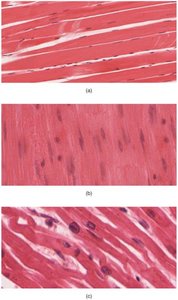
Types of Muscle Tissue
Muscle tissue is a fundamental component of the human body, responsible for movement, posture, and various physiological functions. There are three primary types of muscle tissue:
Skeletal muscle: Voluntary, striated muscle attached to bones, responsible for body movement.
Cardiac muscle: Involuntary, striated muscle found only in the heart, responsible for pumping blood.
Smooth muscle: Involuntary, non-striated muscle found in walls of internal organs, responsible for movements such as peristalsis.
Functions of Skeletal Muscle
Skeletal muscle plays a crucial role in voluntary movements and maintaining homeostasis:
Movement: Contraction of skeletal muscles enables locomotion and manipulation of the environment.
Posture: Stabilizes body position and maintains posture.
Temperature regulation: Generates heat during contraction, helping maintain body temperature.
Voluntary control: Allows conscious control over swallowing, defecation, and urination.
Support: Supports soft tissues, such as the abdominal wall.
Structure of Skeletal Muscle
Connective Tissue Layers
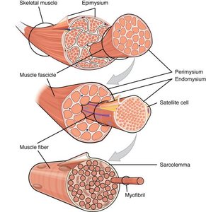
Skeletal muscle is organized into layers of connective tissue that provide structure and support:
Epimysium: Outermost layer, dense collagen fibers, surrounds the entire muscle and separates it from other tissues.
Perimysium: Surrounds bundles of muscle fibers called fascicles, dividing the muscle into compartments.
Endomysium: Encases individual muscle fibers within a fascicle.
Satellite cells: Stem cells involved in muscle repair, located between the endomysium and muscle fibers.
Vascular and Nervous Supply
The epimysium and perimysium contain blood vessels and nerves that supply muscle fibers. Capillary networks in the endomysium ensure nutrient and oxygen delivery and waste removal.
Cellular Structure of Skeletal Muscle
Skeletal muscle fibers are large, multinucleate cells specialized for contraction:
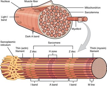
Sarcolemma: Plasma membrane of the muscle fiber.
Sarcoplasm: Cytoplasm of the muscle fiber.
Sarcoplasmic reticulum (SR): Specialized smooth endoplasmic reticulum for calcium storage.
Myofibrils: Cylindrical structures composed of bundles of myofilaments (actin and myosin).
Organization of Myofibrils and Sarcomeres
Myofilaments and Sarcomeres
Myofilaments are organized into repeating units called sarcomeres, the functional unit of muscle contraction:
Thin filaments: Composed primarily of actin.
Thick filaments: Composed primarily of myosin.
Regulatory proteins: Troponin and tropomyosin regulate contraction.
Striated Appearance
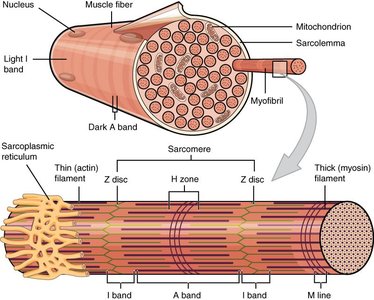
The arrangement of actin and myosin filaments gives skeletal muscle its characteristic striated (banded) appearance.
Neuromuscular Junction and Muscle Activation
Neuromuscular Junction (NMJ)
The NMJ is the site where a motor neuron communicates with a muscle fiber to initiate contraction:
Each muscle fiber is innervated by a motor neuron at the NMJ.
Excitation signals from the neuron are required for muscle contraction.
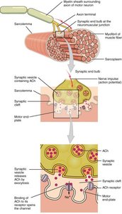
Membrane Potential and Action Potential
Muscle fibers have a resting membrane potential. Neurons and muscle cells use this electrical gradient to generate action potentials, which are essential for muscle contraction.
Excitation-Contraction Coupling
For contraction to occur, the muscle fiber must be stimulated to fire an action potential:
Neuronal action potential travels to the NMJ.
Acetylcholine (ACh) is released and binds to receptors on the motor end-plate.
Na+ ions enter the muscle fiber, generating a muscle action potential.
The action potential spreads along the sarcolemma and into T-tubules, triggering Ca2+ release from the SR.
Mechanism of Muscle Contraction
Role of Calcium and Regulatory Proteins
Calcium ions released from the SR initiate contraction by interacting with regulatory proteins:
Troponin binds calcium, changing shape and moving tropomyosin off actin's active sites.
Myosin heads bind to actin, forming cross-bridges.
Cross-Bridge Cycle and ATP
The cross-bridge cycle is the fundamental process of muscle contraction:
Myosin heads are cocked and bind to actin.
ADP and Pi are released, and the myosin head pivots (power stroke), pulling actin filaments.
ATP binds to myosin, causing detachment from actin.
ATP is hydrolyzed, re-cocking the myosin head for another cycle.
Sliding Filament Model
Muscle contraction shortens the sarcomere as actin filaments slide past myosin filaments:
Occurs in every sarcomere along the myofibril.
Results in shortening of the muscle fiber and overall muscle contraction.
Energy for Muscle Contraction
ATP Sources
ATP is essential for both contraction and relaxation. Muscle fibers have limited ATP stores and rely on three pathways for ATP generation:
Creatine phosphate: Provides energy for ~15 seconds of contraction.
Glycolysis: Anaerobic breakdown of glucose, produces 2 ATP per glucose.
Aerobic respiration: Occurs in mitochondria, produces ~34 ATP per glucose (via pyruvate).
Glycolysis and Aerobic Respiration
During intense exercise, when oxygen is limited, glycolysis becomes the primary source of ATP. Pyruvate is converted to lactic acid, which is transported to the liver and converted back to glucose (Cori Cycle).
Summary Table: Muscle Tissue Types
Type | Location | Control | Appearance |
|---|---|---|---|
Skeletal | Attached to bones | Voluntary | Striated |
Cardiac | Heart | Involuntary | Striated |
Smooth | Walls of organs | Involuntary | Non-striated |
Key Terms and Definitions
Sarcomere: Functional unit of muscle contraction.
Myofibril: Bundle of myofilaments within a muscle fiber.
Epimysium: Outermost connective tissue layer.
Perimysium: Surrounds fascicles.
Endomysium: Surrounds individual muscle fibers.
Neuromuscular junction: Site of communication between motor neuron and muscle fiber.
Creatine phosphate: Molecule that stores energy for rapid ATP regeneration.
Glycolysis: Anaerobic process producing ATP and pyruvate from glucose.
Aerobic respiration: Oxygen-dependent process producing large amounts of ATP.
Important Equations
ATP hydrolysis (energy release):
Glycolysis (per glucose):
Aerobic respiration (per pyruvate):
Sliding filament model:
Additional info: Academic context was added to clarify the mechanisms of muscle contraction, energy pathways, and structural organization. Images were included only when directly relevant to the explanation of muscle tissue structure and function.
