 뒤로
LEC 4
스터디 가이드 - 스마트 노트
 자료에 맞춘 맞춤형 노트, 핵심 정의, 예시, 맥락을 확장해 제공합니다.
Muscular System Overview
Introduction to the Muscular System
The muscular system is essential for movement, support, and heat production in the human body. It is composed of various muscle types and connective tissues that work together to facilitate locomotion and maintain posture.
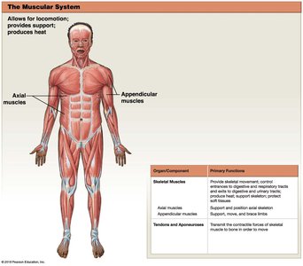
Skeletal muscles are responsible for voluntary movements and are attached to bones.
Axial muscles position the head, neck, and vertebral column, and support respiration.
Appendicular muscles stabilize or move the appendicular skeleton and support fine motor skills.
Muscle Diversity and Representation
Anatomy textbooks often present a limited perspective, typically featuring models of white males and not representing the diversity of body shapes and anatomical variations. It is important to recognize that "normal" anatomy can vary significantly among individuals.
Muscle Tissue Types
Classification of Muscle Tissue
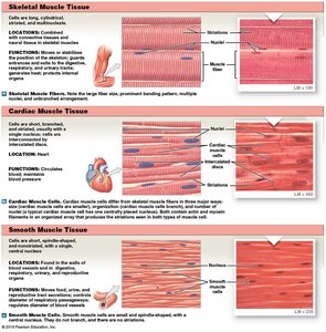
There are three primary types of muscle tissue, each with distinct structural and functional characteristics.
Skeletal muscle: Long, cylindrical, striated cells; voluntary control; attached to bones for movement.
Cardiac muscle: Branched, striated cells; involuntary control; found only in the heart.
Smooth muscle: Spindle-shaped, non-striated cells; involuntary control; found in walls of internal organs.
Gross Anatomy of Skeletal Muscle
Muscle Structure and Organization
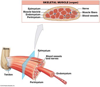
Skeletal muscle is organized into hierarchical layers of connective tissue that surround muscle fibers and fascicles.
Endomysium: Surrounds individual muscle fibers.
Perimysium: Surrounds bundles of muscle fibers (fascicles).
Epimysium: Surrounds the entire muscle.
Tendon: Connects muscle to bone; ligament connects bone to bone.
Muscle Shape and Fiber Arrangement
Classification by Shape and Fiber Arrangement
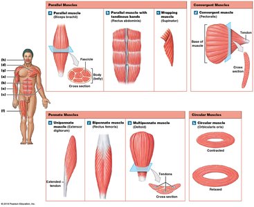
Muscles can be classified based on their shape and the arrangement of their fibers, which influences their function and direction of action.
Parallel muscles: Fibers run parallel to the long axis; contract to shorten and thicken.
Convergent muscles: Fibers converge to a single tendon; allow versatile movement but weaker strength.
Pennate muscles: Fibers attach obliquely to the tendon; can be unipennate, bipennate, or multipennate.
Circular muscles: Fibers arranged in rings; contraction changes diameter of the opening.
Microscopic Anatomy of Skeletal Muscle
Muscle Fiber Structure
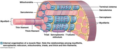
Muscle fibers are specialized cells containing numerous myofibrils, which are responsible for contraction.
Sarcoplasm: Cytoplasm of muscle cell, contains myofibrils.
Transverse (T) tubules: Extensions of the sarcolemma that transmit electrical signals.
Sarcoplasmic reticulum: Stores calcium ions necessary for contraction.
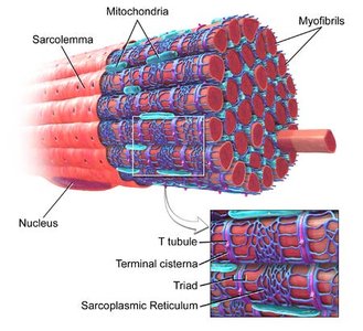
Sarcomere Structure
The sarcomere is the functional contractile unit of muscle, defined by the region between two Z lines.
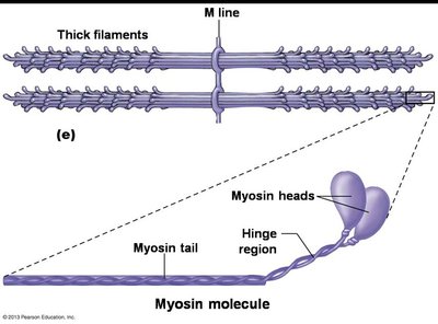
Myosin: Forms thick filaments.
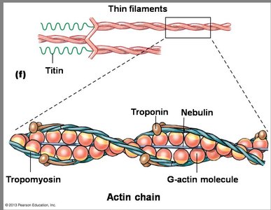
Actin: Forms thin filaments.
Striations: Result from the arrangement of myosin and actin; alternating A-bands (dark) and I-bands (light).
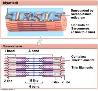
Thick and Thin Filaments
Myosin heads bind to actin during contraction.
Actin filaments are regulated by tropomyosin and troponin.
Muscle Contraction Mechanism
Cross-Bridge Formation and Cycle
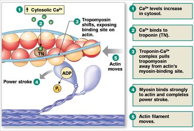
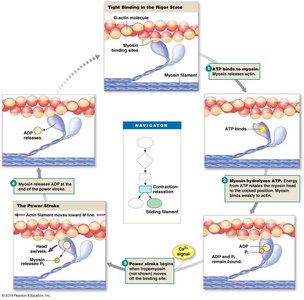
Muscle contraction occurs through the interaction of myosin and actin, regulated by calcium ions and ATP.
Calcium binds to troponin, shifting tropomyosin and exposing binding sites on actin.
Myosin heads bind to actin, forming cross-bridges and pulling the filaments.
ATP is required for myosin head detachment and re-cocking.
Role of Calcium and T-Tubules
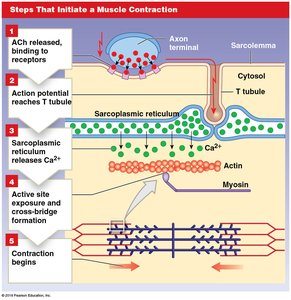
Electrical signals travel through T-tubules to the sarcoplasmic reticulum.
Calcium is released into the sarcoplasm, initiating contraction.
Neuromuscular Junction (NMJ)
Structure and Function of NMJ
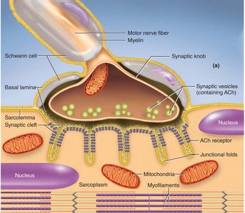
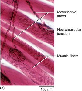
The neuromuscular junction is the site where motor neurons communicate with muscle fibers to initiate contraction.
Acetylcholine (ACh) is released from the axon terminal and binds to receptors on the sarcolemma.
The electrical signal propagates along the sarcolemma and into T-tubules.
Muscle Tension and Motor Units
Muscle Tension and Sarcomere Length
The amount of tension a muscle can generate depends on the overlap between thick and thin filaments in the sarcomere.
Maximum tension occurs when there is optimal overlap.
No tension is produced if filaments are overstretched or compressed.
Motor Units and Recruitment
Motor unit: A motor neuron and all the muscle fibers it innervates.
Small motor units control precise movements; large units control powerful movements.
Recruitment: Activation of additional motor units increases force.
Fast fibers (white muscle) use anaerobic metabolism; slow fibers (red muscle) use aerobic metabolism.
Axial and Appendicular Musculature
Axial Musculature
Positions the head, neck, and vertebral column.
Includes muscles of respiration and core stabilization.
Appendicular Musculature
Stabilizes or moves the appendicular skeleton.
Supports fine motor skills and movement of limbs.
Summary Table: Muscle Types and Functions
Muscle Type | Structure | Location | Function |
|---|---|---|---|
Skeletal | Striated, cylindrical | Attached to bones | Voluntary movement |
Cardiac | Striated, branched | Heart | Pumps blood |
Smooth | Non-striated, spindle-shaped | Walls of organs | Involuntary movement |
Key Terms and Definitions
Endomysium: Connective tissue surrounding each muscle fiber.
Perimysium: Connective tissue surrounding fascicles.
Epimysium: Connective tissue surrounding the entire muscle.
Sarcomere: Functional unit of muscle contraction.
Myofibril: Bundle of contractile proteins within muscle fiber.
Motor unit: Motor neuron and all innervated muscle fibers.
Important Equations
Muscle Force Generation
Muscle force is proportional to the number of cross-bridges formed:
ATP Hydrolysis in Muscle Contraction
ATP is hydrolyzed to provide energy for myosin head movement:
Example: Muscle Contraction Sequence
Action potential arrives at NMJ.
ACh is released and binds to receptors.
Electrical signal travels along sarcolemma and T-tubules.
Sarcoplasmic reticulum releases calcium.
Calcium binds to troponin, exposing actin binding sites.
Myosin binds to actin, forming cross-bridges.
Muscle contracts.
Additional info:
Muscle contraction stops when ACh is broken down and calcium is reabsorbed into the sarcoplasmic reticulum.
Muscle fatigue occurs when ATP supply is depleted or metabolic waste accumulates.
