 뒤로
Nervous System Cells: Structure, Function, and Communication
스터디 가이드 - 스마트 노트
 자료에 맞춘 맞춤형 노트, 핵심 정의, 예시, 맥락을 확장해 제공합니다.
Nervous System Cells
Overview of Nervous System Cells
The nervous system is composed of specialized cells that facilitate rapid communication and integration throughout the body. These cells are classified into two main types: neurons and neuroglia (glial cells).
Neurons: Excitable cells responsible for initiating and conducting electrical impulses. They consist of dendrites, a cell body (soma), and an axon.
Neuroglia: Supportive cells that maintain the environment for neurons, provide insulation, and facilitate efficient functioning.
Cerebrospinal fluid (CSF) is a fluid that circulates through the CNS, providing protection and nutrient transport. The blood-brain barrier (BBB) separates the CNS from the rest of the body, regulating the passage of substances.
Cell bodies of neurons are often clustered together based on function or direction of travel. In the PNS, these clusters are called ganglia; in the CNS, they are called nuclei.
Glial Cells of the Nervous System
Glial cells are essential for supporting neurons and maintaining homeostasis in the nervous system. They differ between the CNS and PNS.
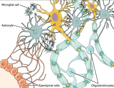
Microglia (CNS): Small, stationary cells that act as macrophages, ingesting pathogens and damaged cells.
Astrocytes (CNS): Star-shaped cells that form tight sheaths around capillaries, contribute to the BBB, and regulate extracellular chemical concentrations.
Ependymal cells (CNS): Epithelial cells lining ventricles, producing and circulating CSF.
Oligodendrocytes (CNS): Cells that wrap around axons to form myelin sheaths, insulating multiple axon segments.
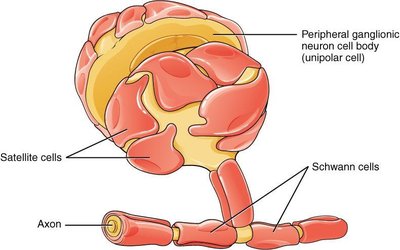
Satellite cells (PNS): Surround neuron cell bodies in ganglia, providing support (similar to astrocytes).
Schwann cells (PNS): Insulate axons with myelin, wrapping around a single axon segment.
Myelin and Myelination
Myelin is a lipid-rich insulation formed by glial cells around axons, increasing the speed of electrical signal transmission. In the PNS, Schwann cells myelinate individual axon segments, while in the CNS, oligodendrocytes can myelinate multiple segments.
Myelin increases axon diameter and gives a white appearance to axons (white matter).
Myelination involves wrapping the cell membrane multiple times around the axon.
Nodes of Ranvier are gaps in the myelin sheath that facilitate rapid signal conduction.
The Neuron
Parts of a Neuron
Neurons are highly specialized cells with distinct structural regions that facilitate the flow of information.
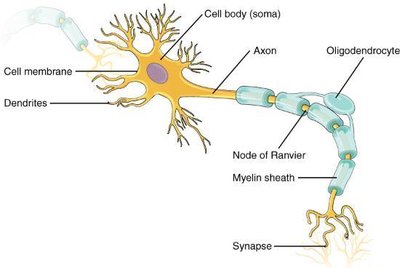
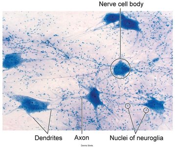
Cell body (soma): Contains the nucleus and major organelles.
Cell membrane (neurolemma): Surrounds the neuron.
Dendrites: Branched processes that receive input from other neurons.
Axon: Long process that transmits impulses to target cells.
Axon hillock: Region where the axon emerges from the soma.
Node of Ranvier: Gaps in myelin sheath affecting signal speed.
Axon terminal: End of axon, branches toward target cell.
Synapse: Specialized contact area for communication with other cells.
Information flows from dendrites to the cell body, then down the axon to the synapse.
Classification of Neurons
Neurons are classified based on the number of processes and the direction of information flow.
Unipolar cells: One process splits into dendrites and axon; found in sensory neurons.
Bipolar cells: Two processes (one axon, one dendrite); rare, found in olfactory epithelium and retina.
Multipolar cells: Three or more processes (one axon, multiple dendrites); most common type.
Functional Regions of the Neuron
Neurons have distinct functional zones:
Input zone: Dendrites and cell body receive signals.
Summation zone: Axon hillock integrates inputs and initiates action potential if threshold is reached.
Conduction zone: Axon transmits action potential.
Output zone: Axon terminals release neurotransmitters to communicate with effector cells.
Synapses and Neurotransmission
Types of Synapses
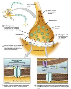
Synapses are junctions where nerve signals are transmitted from a neuron to its effector cell (another neuron, muscle, or gland).
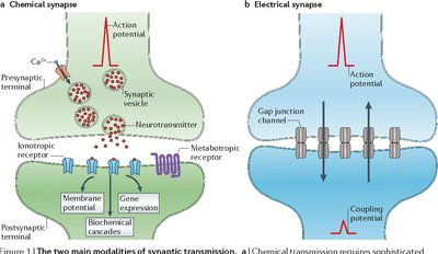
Electrical synapse: Found in cardiac and smooth muscle; action potential spreads via ions through gap junctions.
Chemical synapse: More common; neurotransmitter released by neuron binds to specific receptor on effector cell, causing a response.
Components of a Chemical Synapse
Presynaptic neuron: Releases neurotransmitter.
Synaptic cleft: Space between presynaptic neuron and effector cell.
Neurotransmitter: Chemical messenger released into the synaptic cleft.
Synaptic vesicles: Contain neurotransmitter.
Postsynaptic (effector) cell: Receives signal.
Postsynaptic receptors: Bind neurotransmitter and initiate cellular response.
Neurotransmitter Trafficking
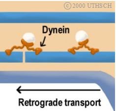
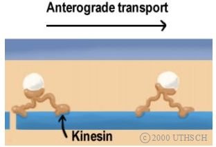
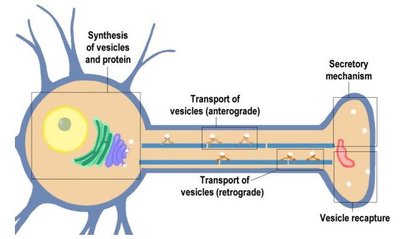
Neurotransmitter vesicles are transported within neurons by motor proteins along microtubules.
Anterograde transport: Kinesin moves vesicles from cell body to axon terminal.
Retrograde transport: Dynein moves vesicles from axon terminal back to cell body.
Role of Neurotransmitter at the Synapse
Neurotransmitters act briefly at the synapse and are terminated by several mechanisms:
Reuptake: Neurotransmitter is transported back into the presynaptic neuron.
Enzymatic degradation: Neurotransmitter is broken down by enzymes.
Diffusion: Neurotransmitter diffuses away and is taken up by glial cells.
Neurotransmitter Receptors
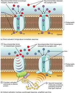
Neurotransmitters bind to specific receptors on the postsynaptic membrane, causing effects via two main receptor types:
Ionotropic receptors: Ligand-gated ion channels; fast response, direct opening of ion channels (e.g., nicotinic acetylcholine receptor).
Metabotropic receptors: G protein-coupled; slower response, can cause opening/closing of ion channels, protein synthesis, or enzyme activation.
Neurotransmitter Systems
Different neurotransmitter systems use specific neurotransmitters, receptors, and mechanisms for action termination. Their effects can be excitatory (depolarization) or inhibitory (hyperpolarization).
System | Example Neurotransmitters | Receptors | Termination | Postsynaptic Effect |
|---|---|---|---|---|
Cholinergic | Acetylcholine | Nicotinic, muscarinic | Degradation by acetylcholinesterase | Nicotinic: depolarization; Muscarinic: variable |
Amino acids | Glutamate, glycine, GABA | Glu, gly, GABA | Reuptake by neurons or glia | Glu: depolarization; Gly/GABA: hyperpolarization |
Biogenic amines | Serotonin, dopamine, norepinephrine, epinephrine | 5-HT, D1/D2, α/β-adrenergic | Reuptake by neurons | Various: depolarization, hyperpolarization, gene transcription, metabolic processes |
Neuropeptides | Met-enkephalin, beta-endorphin, VIP, Substance P | Specific to peptides | Degradation by peptidases | Depolarization or hyperpolarization |
Summary
The nervous system relies on the coordinated function of neurons and glial cells to transmit, process, and integrate information. Myelination, synaptic transmission, and neurotransmitter systems are fundamental to rapid and precise communication within the body.
