 뒤로
Nervous System Composition and Physiology: Mini-Textbook Study Notes
스터디 가이드 - 스마트 노트
 자료에 맞춘 맞춤형 노트, 핵심 정의, 예시, 맥락을 확장해 제공합니다.
Nervous System Composition
Overview of the Nervous System
The nervous system is a complex network responsible for coordinating bodily functions and enabling communication between different parts of the body. It is divided into the central nervous system (CNS) and peripheral nervous system (PNS), each with distinct roles and components.
CNS: Composed of the brain and spinal cord; processes and integrates information.
PNS: Consists of nerves and ganglia outside the CNS; transmits signals to and from the CNS.
Enteric Nervous System: Controls gastrointestinal function independently but communicates with CNS.
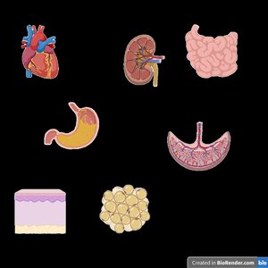
Branches and Pathways of the Peripheral Nervous System
The PNS is further divided into afferent (sensory) and efferent (motor) pathways, which are responsible for transmitting information to and from the CNS.
Afferent Pathways: Carry sensory information from receptors to the CNS.
Efferent Pathways: Transmit motor commands from the CNS to effectors (muscles and glands).
Somatic Nervous System: Controls voluntary movements via skeletal muscles.
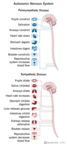
Autonomic Nervous System: Regulates involuntary functions; divided into sympathetic and parasympathetic divisions.
Anatomy of a Peripheral Neuron
Neurons are the fundamental units of the nervous system, specialized for transmitting electrical and chemical signals.
Soma: Cell body containing the nucleus and most organelles.
Dendrites: Receive incoming signals.
Axon: Conducts electrical impulses (action potentials).
Axon Hillock: Initiates action potentials.
Axon Terminals: Release neurotransmitters to communicate with other cells.
Axoplasmic Transport
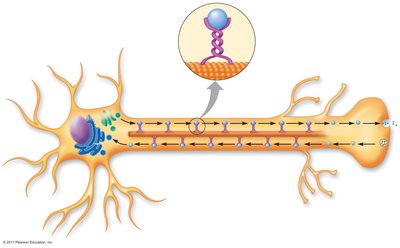
Axoplasmic transport is the process by which materials are moved along the axon, essential for neuronal function and survival.
Anterograde Transport: Moves materials away from the soma toward axon terminals (mediated by kinesin).
Retrograde Transport: Moves materials toward the soma (mediated by dynein).
Synaptic Vesicles: Carry neurotransmitters for release at synapses.
Cells of the Nervous System
Neurons and Glial Cells
The nervous system contains two main cell types: neurons (excitable cells) and glial cells (support cells).
Neurons: Make up about 10% of nervous tissue; responsible for signal transmission.
Glial Cells: Compose 75–90% of CNS; provide structural and functional support.
Types of Glial Cells
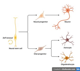
Glial cells are essential for maintaining homeostasis, protecting neurons, and facilitating signal transmission.
Astrocytes: Regulate homeostasis, form the blood-brain barrier, modulate synaptic activity, and protect neurons.
Ependymal Cells: Line CNS cavities and produce cerebrospinal fluid (CSF).
Microglia: Act as resident immune cells, performing phagocytosis and protecting against oxidative stress.
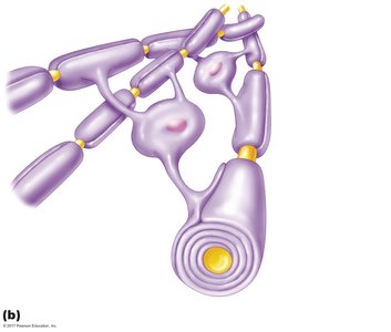
Oligodendrocytes: Form myelin in the CNS.
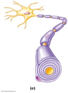
Schwann Cells: Form myelin in the PNS.
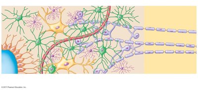
Myelin Sheath-Forming Glia
Myelin is a lipid-rich substance that insulates axons, increasing the speed of electrical signal transmission.
Oligodendrocytes: Myelinate multiple axons in the CNS.
Schwann Cells: Myelinate single axons in the PNS.
Node of Ranvier: Gaps in myelin sheath where action potentials are regenerated.
Protective Structures of the CNS
Meninges
The CNS is protected by three layers of connective tissue called meninges.
Dura Mater: Tough, outermost layer.
Arachnoid Mater: Middle, web-like layer.
Pia Mater: Delicate, innermost layer.
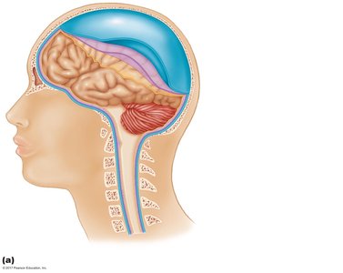
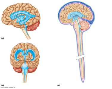
Cerebrospinal Fluid (CSF) and Ventricular System
CSF is a clear fluid that bathes the CNS, providing physical and metabolic support.
Produced by: Ependymal cells in the choroid plexus.
Circulation: Flows through ventricles and subarachnoid space; reabsorbed by arachnoid villi.
Functions: Cushions the brain, maintains stable interstitial environment.
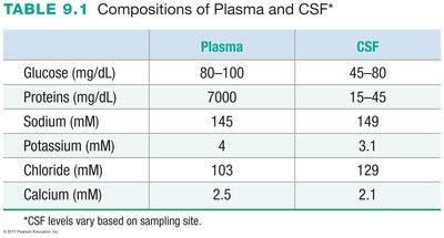
Comparison of Plasma and CSF Composition
Plasma and CSF differ in their chemical composition, reflecting their distinct roles in the body.
Component | Plasma | CSF |
|---|---|---|
Glucose (mg/dL) | 80–100 | 45–80 |
Proteins (mg/dL) | 7000 | 15–45 |
Sodium (mM) | 145 | 149 |
Potassium (mM) | 4 | 3.1 |
Chloride (mM) | 103 | 129 |
Calcium (mM) | 2.5 | 2.1 |
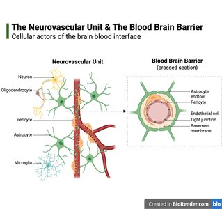
Blood-Brain Barrier and Neurovascular Unit
Blood-Brain Barrier (BBB)
The BBB is a selective barrier formed by endothelial cells with tight junctions, astrocyte end-feet, and pericytes. It regulates the exchange of substances between blood and CNS, maintaining homeostasis and protecting against toxins and pathogens.
Capillaries: Non-fenestrated, contain tight junctions.
Functions: Restricts passage of pathogens, toxins, and large molecules; selectively transports nutrients; clears waste.
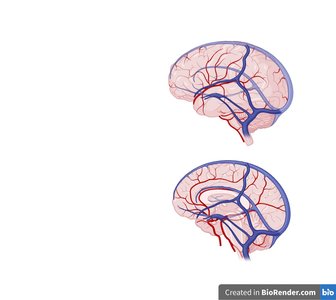
Blood Supply to the CNS
Metabolic Demands and Stroke
The CNS has high metabolic demands, requiring continuous blood supply for oxygen and glucose. Disruption of blood flow can lead to stroke, resulting in neuronal injury or death.
Brain Weight: 3–4 pounds (~2% of body weight).
Blood Supply: Receives 15% of cardiac output.
Oxygen and Glucose Use: 20% and 50% of body consumption at rest, respectively.
Stroke: Caused by occlusion or hemorrhage of cerebral blood vessels.
Transient Ischemic Attack (TIA): Temporary reduction in blood supply (“mini-stroke”).
Types of Stroke
Stroke can be classified based on its underlying mechanism.
Atherosclerotic Stroke: Blockage due to plaque buildup.
Hemorrhagic Stroke: Rupture and bleeding of blood vessels.
Ischemic Stroke: Blockage due to thrombus formation.
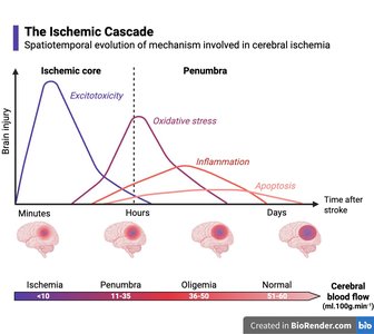
Ischemic Cascade and Brain Injury
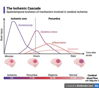
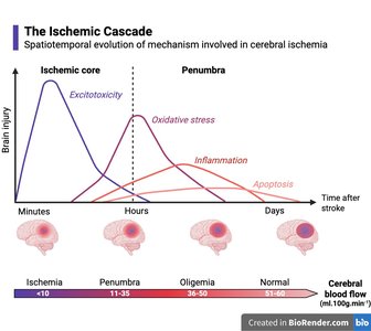
The ischemic cascade describes the sequence of events following reduced blood flow to the brain, leading to excitotoxicity, oxidative stress, inflammation, and apoptosis.
Ischemic Core: Area of irreversible damage.
Penumbra: Area of potentially reversible injury.
Oligemia: Reduced blood flow but not severe enough to cause cell death.
Summary Table: Nervous System Components
Component | Function |
|---|---|
Neuron | Signal transmission |
Astrocyte | Homeostasis, BBB formation |
Microglia | Immune defense |
Oligodendrocyte | Myelination in CNS |
Schwann Cell | Myelination in PNS |
Ependymal Cell | CSF production |
Key Equations
Action Potential Propagation
Membrane potential change:
Nernst equation for ion equilibrium:
Additional info:
Some context and explanations were expanded for clarity and completeness.
Tables and images were recreated and placed only where directly relevant to the content.
