 뒤로
Nervous Tissue and Physiology: Structure and Function
스터디 가이드 - 스마트 노트
 자료에 맞춘 맞춤형 노트, 핵심 정의, 예시, 맥락을 확장해 제공합니다.
Nervous Tissue and Physiology
Overview of the Nervous System
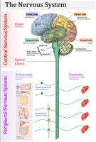
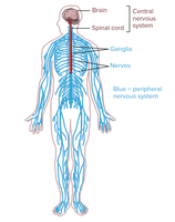
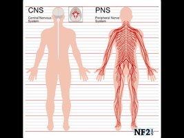
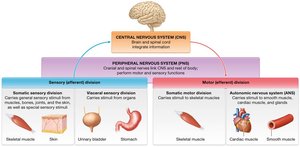
The nervous system is a physically connected network of cells, tissues, and organs that enables communication, environmental response, and the performance of life activities. It is divided into two main anatomical subdivisions: the Central Nervous System (CNS) and the Peripheral Nervous System (PNS).
Central Nervous System (CNS): Consists of the brain and spinal cord; serves as the integrating and control center.
Peripheral Nervous System (PNS): Composed of cranial nerves, spinal nerves, and their branches; links the CNS to the rest of the body.
Organs of the Nervous System
The nervous system is an organ system composed of the following organs:
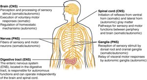
Brain: Responsible for perception, processing of sensory stimuli, voluntary motor control, and regulation of homeostasis.
Spinal Cord: Initiates reflexes and serves as a pathway for information between the brain and peripheral organs.
Nerves: Bundles of axons, blood vessels, and connective tissue that transmit signals throughout the body.
Ganglia: Collections of neuron cell bodies in the PNS.
Primary Functions of the Nervous System
The nervous system performs three primary functions:
Sensory Input: Gathering information from internal and external environments.
Integration: Interpreting sensory input and determining appropriate responses.
Motor Output: Activating effector organs (muscles and glands) to cause a response.
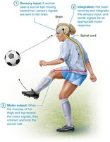
Anatomical and Functional Subdivisions
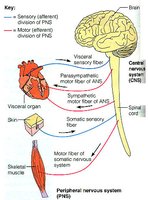
The nervous system is further divided into functional subdivisions:
Somatic Nervous System: Controls voluntary movements via skeletal muscles.
Autonomic Nervous System (ANS): Regulates involuntary functions in smooth muscle, cardiac muscle, and glands. Includes sympathetic and parasympathetic divisions.
Nervous Tissue Structure
Components of Nervous Tissue
Nervous tissue consists of neurons, neuroglia, and extracellular matrix (ECM). Neurons are excitable cells responsible for transmitting signals, while neuroglia support and protect neurons.
Neurons: Make up 10% of nervous tissue cells; specialized for communication.
Neuroglia: Comprise 90% of nervous tissue cells; provide structural and metabolic support.
Extracellular Matrix: Ground substance and glycoproteins, accounting for 20% of tissue volume.
Structure of a Typical Neuron
Neurons have specialized structures that support their function:
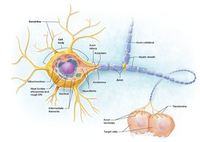
Cell Body (Soma): Contains the nucleus, cytoplasm, mitochondria, Nissl bodies (ribosomes and rough ER), Golgi apparatus, and neurofibrils. Responsible for protein synthesis and metabolic maintenance.
Dendrites: Numerous, short, highly branched processes that receive signals and generate local potentials.
Axon: Single, long process that conducts action potentials. Features include axon hillock, axon collaterals, telodendria, and synaptic knobs. Often wrapped in a myelin sheath for insulation and increased transmission speed.
Neuron Processes and Myelination
Neurons have processes (dendrites and axons) that are organized differently in the CNS and PNS:
CNS: Bundles of processes are called tracts.
PNS: Bundles of processes are called nerves.
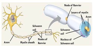
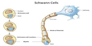
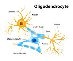
Myelin Sheath: Protein-lipoid extension of glial plasma membrane; acts as insulation and increases transmission speed.
Nodes of Ranvier: Gaps between myelinated segments that facilitate rapid signal transmission.
Axonal Transport
Axonal transport is the movement of materials along the axon:
Slow Axonal Transport: Anterograde only; moves cytoskeletal and other proteins at 1-3 mm/day.
Fast Axonal Transport: Anterograde and retrograde; moves vesicles and organelles at 200-400 mm/day.
Motor Proteins: Kinesin (anterograde) and dynein (retrograde) use ATP for transport.
Classification of Neurons
Structural Classification
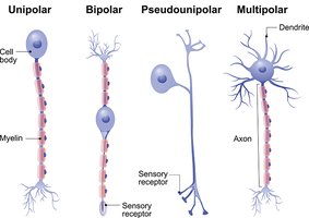
Neurons are classified based on the number and arrangement of their processes:
Multipolar: One axon, many dendrites; most common type.
Bipolar: One axon, one dendrite; rare, found in the retina.
Unipolar: Single process; "T" shaped, found primarily in ganglia of the PNS.
Functional Classification
Neurons are also classified by their function:
Sensory Neurons: Transmit impulses from receptors to CNS; mostly unipolar, cell bodies outside CNS.
Motor Neurons: Carry impulses away from CNS; cell bodies located in CNS.
Interneurons: Connect sensory and motor neurons; primarily multipolar, confined to CNS.
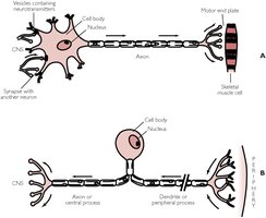
Organization of Neurons in CNS and PNS
Neurons in common signaling pathways group together:
CNS: Cell bodies form nuclei; axons form tracts.
PNS: Cell bodies form ganglia; axons form nerves.
Neuroglia: Support Cells of the Nervous System
Types and Functions of Neuroglia
Neuroglia are essential for maintaining the environment, supporting neurons, and repairing tissue. There are six types:
CNS Neuroglia: Astrocytes, oligodendrocytes, microglial cells, ependymal cells.
PNS Neuroglia: Schwann cells, satellite cells.
CNS Neuroglia
Astrocytes: Anchor neurons and blood vessels, maintain extracellular environment, assist in blood-brain barrier formation, repair damaged tissue.
Oligodendrocytes: Form myelin sheaths around CNS axons.
Microglial Cells: Act as macrophages, phagocytize foreign invaders, monitor neuron health.
Ependymal Cells: Line brain ventricles, produce and circulate cerebrospinal fluid.
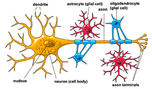
PNS Neuroglia
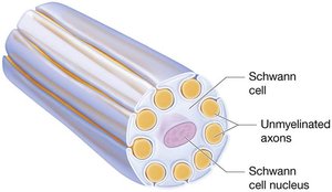
Schwann Cells: Form myelin sheaths around PNS axons.
Satellite Cells: Surround and support neuron cell bodies in ganglia.
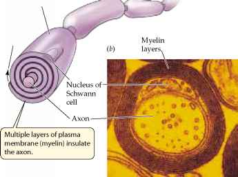
Myelination in CNS and PNS
Myelin Sheath Structure and Function
Myelin sheaths are electrical insulators composed of multiple phospholipid bilayers. They increase the speed of nerve impulse transmission.
CNS: Oligodendrocytes wrap around many axons; myelination begins after birth; no neurolemma.
PNS: Schwann cells wrap around part of a single axon; myelination begins in early fetal period; outer layer forms neurolemma.
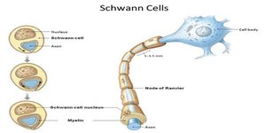
White Matter and Gray Matter
White Matter: Composed of myelinated axons; appears white.
Gray Matter: Composed of neuron cell bodies, unmyelinated dendrites, and axons; appears gray.
Axon Repair and Regeneration
Repair Mechanisms
Axon repair differs between the CNS and PNS:
CNS: Astrocytes form scar tissue; damaged neurons cannot be replaced.
PNS: Axon regeneration is possible if the cell body and part of the myelinated axon are present. Schwann cells and basal lamina form a regeneration tube to guide regrowth.
Steps of Axon Repair in PNS
Wallerian degeneration: Phagocytes digest components distal to severance point.
Growth processes form: Several proximal cell extensions sprout from axon connected to cell body.
Regeneration tube forms: Schwann cells and basal lamina form a regeneration tube.
Regeneration tube directs growth: Growth process in tube stimulated to continue growing.
Connection with target cell reestablished: Schwann cells form myelin sheaths, muscle atrophy halted.
Note: Neurons, once formed, cannot undergo mitosis due to loss of centrioles.
