 뒤로
Neural Tissue: Structure, Function, and Clinical Relevance
스터디 가이드 - 스마트 노트
 자료에 맞춘 맞춤형 노트, 핵심 정의, 예시, 맥락을 확장해 제공합니다.
Neural Tissue and the Nervous System
Overview of the Nervous System
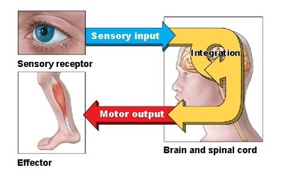
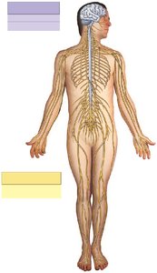
The nervous system is a complex network responsible for integrating, processing, and coordinating sensory input and motor output. It is divided into two main anatomical subdivisions: the Central Nervous System (CNS) and the Peripheral Nervous System (PNS).
CNS: Consists of the brain and spinal cord. Responsible for higher functions such as intelligence, memory, learning, and emotion.
PNS: Includes all neural tissue outside the CNS. Facilitates communication between the CNS and peripheral tissues and systems.
Afferent vs. Efferent Divisions
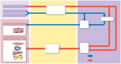
The PNS is functionally divided into afferent (sensory) and efferent (motor) divisions:
Afferent Division: Brings sensory information to the CNS via sensory receptors (e.g., neurons, specialized cells, sense organs).
Efferent Division: Carries motor commands from the CNS to effectors (muscle cells, glands, specialized cells).
Somatic vs. Visceral Components
Both afferent and efferent divisions have somatic and visceral components:
Somatic: Relates to skeletal muscles, joints, and skin. The somatic nervous system (SNS) controls voluntary muscle contraction and reflexes.
Visceral: Relates to internal organs (smooth and cardiac muscle, glands). The autonomic nervous system (ANS) regulates involuntary functions and is divided into sympathetic and parasympathetic divisions.
Neural Tissue: Neurons and Neuroglia
Neurons
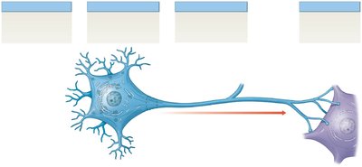
Neurons are the functional cells of the nervous system, responsible for the transfer and processing of information. They are supported by neuroglia (glial cells).
Cell Body: Contains the nucleus and cytoplasm (perikaryon), organelles for energy and biosynthesis. Referred to as "gray matter."
Dendrites: Sensory processes that receive stimuli.
Axon: Long cytoplasmic process (nerve fiber) that propagates action potentials.
Synaptic Terminal: The end of the axon, forms synapses with other neurons or effectors.
Neuroglia
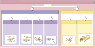
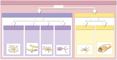
Neuroglia are supporting cells that maintain the environment around neurons, provide structural support, and participate in repair processes. They are classified based on their location:
CNS Neuroglia: Astrocytes, oligodendrocytes, microglia, ependymal cells
PNS Neuroglia: Satellite cells, Schwann cells
Functions of CNS Neuroglia
Astrocytes: Maintain blood-brain barrier, provide structural support, regulate ion/nutrient concentrations, absorb/recycle neurotransmitters, form scar tissue after injury.
Oligodendrocytes: Myelinate CNS axons, provide structural framework.
Microglia: Phagocytic cells that remove debris, wastes, and pathogens.
Ependymal Cells: Line ventricles and central canal, assist in producing and monitoring cerebrospinal fluid (CSF).
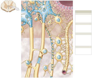
Functions of PNS Neuroglia
Satellite Cells: Surround neuron cell bodies in ganglia, regulate exchange of materials.
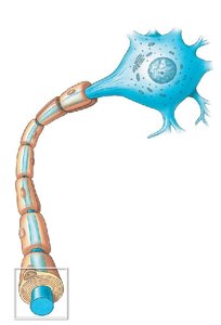
Schwann Cells: Myelinate peripheral axons, participate in repair after injury.
Myelination
Process of Myelination
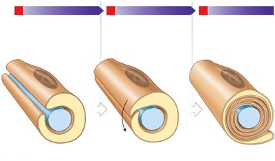
Myelination is the process by which glial cells wrap axons in a myelin sheath, increasing the speed of electrical signal transmission.
Schwann Cells (PNS): Enclose axons, rotate around them, and compress their membranes to form the myelin sheath.
Oligodendrocytes (CNS): Myelinate multiple axons simultaneously.
Classification of Neurons
Structural Classification
Neurons are classified based on the number and arrangement of their processes:
Anaxonic: Small, cannot distinguish dendrites from axons; found in CNS and special sense organs.
Bipolar: One dendrite and one axon; rare, important for sensory information (sight, smell, hearing).
Pseudounipolar: Continuous dendritic and axonal processes; cell body off to one side; usually sensory neurons of PNS.
Multipolar: Several dendrites and a single axon; most common, all motor neurons controlling skeletal muscles.
Functional Classification
Sensory Neurons: Afferent division; deliver information to CNS. Includes exteroceptors (external environment), proprioceptors (position/movement), and interoceptors (internal environment).
Motor Neurons: Efferent division; stimulate or modify activity of peripheral tissues/organs.
Interneurons: Located entirely within CNS; analyze sensory input and coordinate motor output; may be excitatory or inhibitory.
Synapses
Structure and Types of Synapses
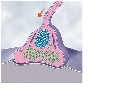
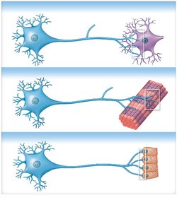
A synapse is the junction where a neuron communicates with another cell. Synapses can occur between neurons, at neuromuscular junctions (with muscle fibers), or at neuroglandular junctions (with gland cells).
Presynaptic Membrane: Releases neurotransmitters.
Postsynaptic Membrane: Receives the signal.
Synaptic Cleft: Gap between the two membranes.
Neuron Processing and Circuits
Types of Neuronal Circuits
Neuronal processing involves the organization of neurons into circuits or pools:
Divergence: One neuron spreads information to several neurons.
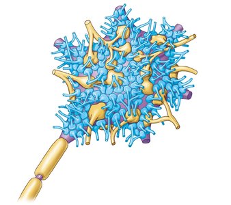
Convergence: Several neurons synapse onto the same postsynaptic neuron.
Serial Processing: Neurons work sequentially in a relay.
Parallel Processing: Several neurons process the same information simultaneously.
Reverberation: Positive feedback circuit for complex control.
Clinical Anatomy
Multiple Sclerosis (MS)
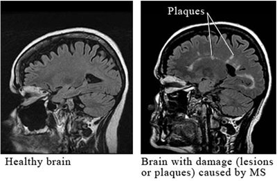
Multiple Sclerosis is an autoimmune disorder of the CNS where the immune system attacks the myelin sheath surrounding nerve fibers, primarily targeting oligodendrocytes. This disrupts electrical signal transmission, leading to a range of symptoms:
Vision problems
Sensory changes
Fatigue and weakness
Mobility and balance issues
Disease-modifying therapies aim to suppress or alter the immune response.
Myasthenia Gravis
Myasthenia Gravis is a clinical condition affecting neuromuscular junctions, leading to muscle weakness. It is caused by an autoimmune response against acetylcholine receptors.
Summary Table: Neuroglia Types and Functions
Location | Type | Function |
|---|---|---|
CNS | Astrocytes | Maintain BBB, structural support, regulate environment, scar tissue |
CNS | Oligodendrocytes | Myelinate axons, structural framework |
CNS | Microglia | Phagocytosis, remove debris/pathogens |
CNS | Ependymal cells | Line ventricles/central canal, produce/monitor CSF |
PNS | Satellite cells | Regulate environment around neuron cell bodies |
PNS | Schwann cells | Myelinate axons, repair after injury |
Key Terms and Definitions
Neuron: Functional cell of the nervous system, transmits electrical signals.
Neuroglia: Supporting cells, maintain environment and provide structural support.
Myelination: Formation of a myelin sheath around axons for faster signal transmission.
Synapse: Junction between two neurons or a neuron and an effector.
Central Nervous System (CNS): Brain and spinal cord.
Peripheral Nervous System (PNS): All neural tissue outside CNS.
Somatic Nervous System (SNS): Controls voluntary muscle movements.
Autonomic Nervous System (ANS): Controls involuntary functions.
Equations and Formulas
Action potential propagation is governed by the movement of ions across the neuronal membrane, described by the Nernst equation:
Where: E = membrane potential R = gas constant T = temperature z = charge of ion F = Faraday's constant [ion outside], [ion inside] = ion concentrations
Additional info: Academic context was added to clarify the functions of neuroglia, neuron structure, and clinical relevance of MS and Myasthenia Gravis.
