 뒤로
Peripheral Nervous System and Reflex Activity: Structure, Function, and Clinical Relevance
스터디 가이드 - 스마트 노트
 자료에 맞춘 맞춤형 노트, 핵심 정의, 예시, 맥락을 확장해 제공합니다.
Principles of Structure and Function of the Peripheral Nervous System (PNS)
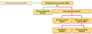
Overview of the Nervous System
The nervous system is divided into the Central Nervous System (CNS) and the Peripheral Nervous System (PNS). The PNS connects the CNS to the rest of the body and is responsible for transmitting sensory and motor information.
Sensory (Afferent) Division: Carries sensory information from receptors to the CNS.
Motor (Efferent) Division: Transmits motor commands from the CNS to effectors (muscles and glands).
Somatic Nervous System: Controls voluntary movements of skeletal muscles.
Autonomic Nervous System (ANS): Regulates involuntary functions; subdivided into Sympathetic and Parasympathetic divisions.
Structural Organization of the PNS
Nerve Structure
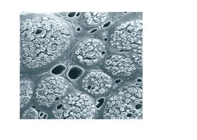
Nerves are bundles of axons grouped into fascicles, each surrounded by connective tissue wrappings:
Epineurium: Outermost layer, encloses the entire nerve.
Perineurium: Surrounds each fascicle.
Endoneurium: Encloses individual axons.
Blood vessels are present within nerves to supply nutrients.
Classification and Function of Peripheral Nerves
Nerve Function
Peripheral nerves are classified based on their function:
Sensory (Afferent): Transmit sensory information to the CNS.
Motor (Efferent): Carry motor commands from the CNS to effectors.
Mixed: Contain both sensory and motor fibers.
Nerve Classification
Cranial Nerves: Originate from the brain; 12 pairs.
Spinal Nerves: Originate from the spinal cord; 31 pairs.
Cranial Nerves: Structure and Function
Olfactory Nerve (CN I)
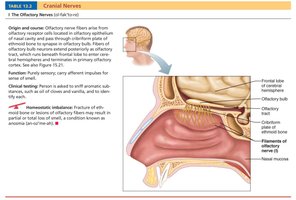
The olfactory nerve is responsible for the sense of smell. Fibers arise from olfactory receptor cells in the nasal cavity and synapse in the olfactory bulb. Sensory information is delivered to the olfactory cortex in the temporal lobe.
Function: Smell (sensory only)
Clinical Relevance: Damage may result in anosmia (loss of smell).
Optic Nerve (CN II)
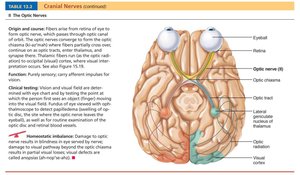
The optic nerve transmits visual information from the retina to the brain. Fibers converge at the optic chiasm, partially cross, and continue as optic tracts to the thalamus and then to the visual cortex in the occipital lobe.
Function: Vision (sensory only)
Clinical Relevance: Damage to the optic nerve or chiasm can cause visual field defects.
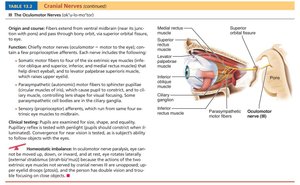
Oculomotor Nerve (CN III)
The oculomotor nerve arises from the midbrain and innervates most of the extrinsic eye muscles. It also carries parasympathetic fibers to the iris and ciliary muscle, controlling pupil constriction and lens shape.
Function: Eye movement, pupil constriction, lens accommodation (motor and some sensory)
Clinical Relevance: Damage can cause double vision, drooping eyelid, and inability to move the eye properly.
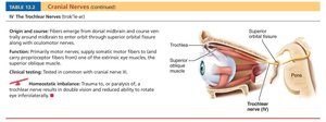
Trochlear Nerve (CN IV)
The trochlear nerve arises from the midbrain and innervates the superior oblique muscle of the eye, enabling downward and lateral movement.
Function: Eye movement (motor and some sensory)
Clinical Relevance: Damage results in difficulty moving the eye downward and laterally.
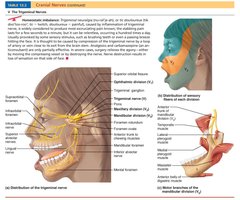
Trigeminal Nerve (CN V)
The trigeminal nerve is a mixed nerve with three divisions: ophthalmic, maxillary, and mandibular. It provides sensory innervation to the face and motor innervation to muscles of mastication.
Function: Sensation of the face, chewing (mixed)
Clinical Relevance: Damage can cause loss of facial sensation or difficulty chewing.
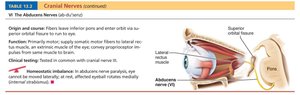
Abducens Nerve (CN VI)
The abducens nerve arises from the pons and innervates the lateral rectus muscle of the eye, enabling lateral movement.
Function: Lateral eye movement (motor and some sensory)
Clinical Relevance: Damage results in inability to move the eye laterally.
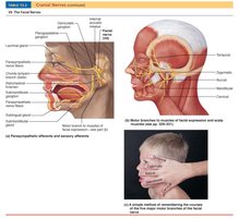
Facial Nerve (CN VII)
The facial nerve arises from the pons and innervates muscles of facial expression, glands for tear and saliva production, and carries taste sensations from the anterior two-thirds of the tongue.
Function: Facial expression, taste, glandular secretion (mixed)
Clinical Relevance: Damage can cause facial paralysis or loss of taste.
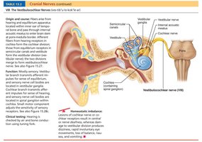
Vestibulocochlear Nerve (CN VIII)
The vestibulocochlear nerve transmits balance and hearing information from the inner ear to the brain.
Function: Hearing and balance (sensory only)
Clinical Relevance: Damage can cause hearing loss or balance disorders.
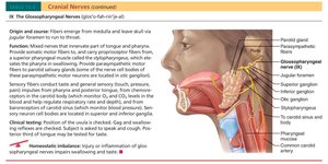
Glossopharyngeal Nerve (CN IX)
The glossopharyngeal nerve arises from the medulla and innervates the tongue and pharynx, contributing to taste, swallowing, and salivary secretion.
Function: Taste, swallowing, salivation (mixed)
Clinical Relevance: Damage can affect swallowing and taste.
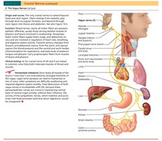
Vagus Nerve (CN X)
The vagus nerve arises from the medulla and innervates thoracic and abdominal viscera, controlling heart rate, digestion, and other autonomic functions.
Function: Autonomic control of viscera, swallowing, taste (mixed)
Clinical Relevance: Damage can affect heart rate, digestion, and swallowing.
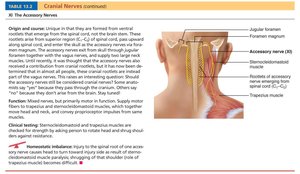
Accessory Nerve (CN XI)
The accessory nerve arises from the spinal cord and innervates the sternocleidomastoid and trapezius muscles, enabling head and shoulder movement.
Function: Head and shoulder movement (motor and some sensory)
Clinical Relevance: Damage can cause weakness in these muscles.
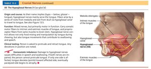
Hypoglossal Nerve (CN XII)
The hypoglossal nerve arises from the medulla and innervates muscles of the tongue, enabling speech and swallowing.
Function: Tongue movement (motor and some sensory)
Clinical Relevance: Damage can cause difficulty speaking or swallowing.
Spinal Nerves and Plexuses
Spinal Nerve Structure
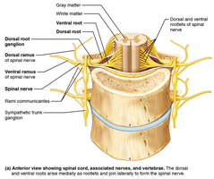
Spinal nerves are mixed nerves formed by the joining of dorsal (sensory) and ventral (motor) roots. Each spinal nerve branches into dorsal and ventral rami.
31 pairs of spinal nerves: All mixed nerves
Dorsal root: Sensory fibers
Ventral root: Motor fibers
Dorsal root ganglia: Contain cell bodies of sensory neurons
Spinal Nerve Plexuses
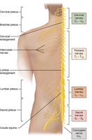
Ventral rami (except T2-T12) form plexuses, interlacing networks that redistribute fibers so each muscle receives neural supply from more than one spinal nerve. The four main plexuses are:
Cervical plexus: C1-C5
Brachial plexus: C5-T1
Lumbar plexus: L1-L4
Sacral plexus: L4-S4
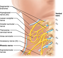
Cervical Plexus
The cervical plexus innervates the neck and diaphragm, including the phrenic nerve (C3-C5), which is essential for breathing.
Summary Table: Cranial Nerves and Their Functions
Cranial Nerve | Function | Type |
|---|---|---|
Olfactory (I) | Smell | Sensory |
Optic (II) | Vision | Sensory |
Oculomotor (III) | Eye movement, pupil constriction | Motor |
Trochlear (IV) | Eye movement | Motor |
Trigeminal (V) | Facial sensation, chewing | Mixed |
Abducens (VI) | Lateral eye movement | Motor |
Facial (VII) | Facial expression, taste | Mixed |
Vestibulocochlear (VIII) | Hearing, balance | Sensory |
Glossopharyngeal (IX) | Taste, swallowing | Mixed |
Vagus (X) | Autonomic control, swallowing | Mixed |
Accessory (XI) | Head/shoulder movement | Motor |
Hypoglossal (XII) | Tongue movement | Motor |
Clinical Relevance
Understanding the structure and function of the PNS and its nerves is essential for diagnosing and treating neurological disorders. Damage to specific nerves can result in loss of sensation, motor function, or autonomic control, depending on the affected region.
Additional info: Academic context was added to clarify nerve functions, clinical relevance, and structural organization. Images were included only when directly relevant to the explanation of the adjacent paragraph.
