 뒤로
Peripheral Nervous System: Structure, Function, and Reflexes
스터디 가이드 - 스마트 노트
 자료에 맞춘 맞춤형 노트, 핵심 정의, 예시, 맥락을 확장해 제공합니다.
Peripheral Nervous System (PNS) and Its Divisions
Overview of PNS Structure and Function
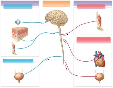
The Peripheral Nervous System (PNS) connects the Central Nervous System (CNS) to limbs and organs, facilitating sensory input and motor output. It is divided into sensory (afferent) and motor (efferent) divisions, each with somatic and visceral components.
Somatic Sensory Division: Carries signals from skin, muscles, bones, joints, and special sensory organs (e.g., eyes).
Visceral Sensory Division: Transmits sensory information from internal organs (viscera).
Somatic Motor Division: Sends motor commands to skeletal muscles.
Visceral Motor Division (Autonomic Nervous System, ANS): Controls involuntary functions in organs such as the heart and urinary bladder.
Cranial Nerves
Structure and Function of Cranial Nerves
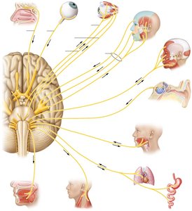
Cranial nerves are twelve pairs of nerves that emerge directly from the brain and brainstem, each with specific sensory, motor, or mixed functions. They innervate the head, neck, and some visceral organs.
CN I (Olfactory): Sense of smell
CN II (Optic): Sense of vision
CN III (Oculomotor): Eye movement, pupil constriction
CN IV (Trochlear): Eye turning
CN V (Trigeminal): Sensation in face, muscles of mastication
CN VI (Abducens): Lateral eye movement
CN VII (Facial): Facial expression, taste (anterior tongue), salivary glands
CN VIII (Vestibulocochlear): Hearing and balance
CN IX (Glossopharyngeal): Taste (posterior tongue), pharynx, parotid gland
CN X (Vagus): Pharynx, tongue, visceral organs
CN XI (Accessory): Neck and shoulder muscles
CN XII (Hypoglossal): Tongue muscles
Spinal Nerves and Plexuses
Classification and Distribution of Spinal Nerves
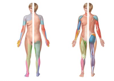
Spinal nerves are classified by their origin in the spinal cord and are organized into plexuses that innervate specific regions of the body. Each plexus contains nerves that serve both sensory and motor functions.
Cervical Plexus (C1–C5): Innervates neck, scalp, and diaphragm.
Brachial Plexus (C5–T1): Innervates upper limb and pectoral girdle.
Lumbar Plexus (L1–L4): Innervates anterior pelvic region and thigh.
Sacral Plexus (L4–S4): Innervates hip and lower leg.
Sensory Receptors and General Senses
Types and Functions of Sensory Receptors
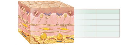
Sensory receptors detect changes in the environment and body, initiating the process of sensation. They are classified by the type of stimulus they detect and their location.
Mechanoreceptors: Detect touch, pressure, vibration, and stretch.
Thermoreceptors: Detect temperature changes.
Nociceptors: Detect pain.
Photoreceptors: Detect light (in the eye).
Chemoreceptors: Detect chemical stimuli (taste, smell).
General senses are distributed throughout the skin, muscles, and joints, while special senses are localized in specific organs.
Receptive Fields and Two-Point Discrimination
Receptive fields are areas served by a particular sensory neuron. Regions with many small receptive fields (e.g., fingertips) have high spatial resolution, while regions with fewer, larger fields (e.g., forearm) have lower resolution. Two-point discrimination threshold measures the ability to distinguish two close stimuli.
Fingertip: Threshold of 3 mm
Forearm: Threshold of 40 mm
Body regions specialized for environmental sensing contain more neurons with smaller receptive fields.
Dermatomes and Clinical Relevance
Dermatomes and Their Mapping
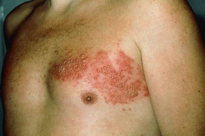
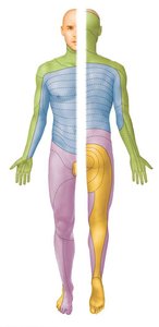
A dermatome is a specific region of skin supplied by a single pair of spinal nerves. Dermatomal maps are used clinically to diagnose nerve or spinal cord injuries.
Referred Pain and Chronic Pain
Mechanism of Referred Pain
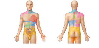
Referred pain occurs when pain is perceived in a region of the body that is not the source of the stimulus. This is common with organ pain, which is often felt in the skin due to convergence of sensory pathways in the spinal cord.
Chronic Pain
Chronic pain is not a direct response to tissue injury and may result from malfunction in the cerebrum or thalamus. It is often associated with psychological symptoms such as depression and frustration.
Neural Reflexes
Reflex Arc and Classification
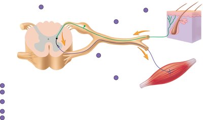
Neural reflexes are rapid, automatic responses to stimuli. The reflex arc consists of five components: sensory receptor, sensory neuron, interneuron (sometimes absent), motor neuron, and effector organ.
Spinal or Cranial: Integration center is spinal cord or brain.
Somatic or Visceral: Effector is skeletal muscle or organ/gland.
Monosynaptic or Polysynaptic: Direct synapse or involves interneurons.
Innate or Acquired: Present at birth or learned.
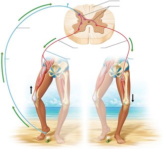
Withdrawal or Crossed-Extensor: Same side or opposite side response.
Flexion (Withdrawal) Reflex and Crossed-Extensor Reflex
The flexion reflex removes a limb from a painful stimulus, while the crossed-extensor reflex maintains balance by extending the opposite limb. Reciprocal innervation ensures relaxation of antagonistic muscles during these reflexes.

Muscle Spindle and Golgi Tendon Organ
Muscle Spindle: Stretch Reflex
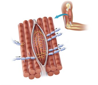
The muscle spindle is a sensory receptor within the muscle that detects stretch and helps maintain muscle tone. The stretch reflex (e.g., patellar reflex) is a simple, monosynaptic reflex that does not involve interneurons.
Muscle spindle: Contains specialized fibers that respond to stretch.
Sensory neurons: Innervate noncontractile centers of spindle cells.
Golgi Tendon Organ: Tendon Reflex
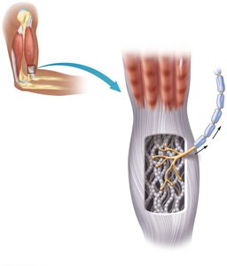
The Golgi tendon organ prevents damage to tendons by inhibiting muscle contraction when excessive tension is detected. This reflex involves inhibitory interneurons and produces sudden muscle relaxation.
Golgi tendon organ: Encapsulated nerve endings associated with collagen fibers in tendons.
Function: Prevents excessive tension and protects tendons.
Summary Table: Cranial Nerves
Number | Name | Function | Related Organs/Stimuli | Path (Foramen) |
|---|---|---|---|---|
CN I | Olfactory | Sensory | Smell | Cribriform plate |
CN II | Optic | Sensory | Vision | Optic canal |
CN III | Oculomotor | Motor | Eye muscles, pupil | Superior orbital fissure |
CN IV | Trochlear | Motor | Eye turning | Superior orbital fissure |
CN V | Trigeminal | Both | Face sensation, mastication | Multiple |
CN VI | Abducens | Motor | Lateral eye movement | Superior orbital fissure |
CN VII | Facial | Both | Facial expression, taste | Internal auditory meatus |
CN VIII | Vestibulocochlear | Sensory | Hearing, balance | Internal auditory meatus |
CN IX | Glossopharyngeal | Both | Taste, pharynx | Jugular foramen |
CN X | Vagus | Both | Pharynx, tongue, viscera | Jugular foramen |
CN XI | Accessory | Motor | Neck, shoulder muscles | Jugular foramen |
CN XII | Hypoglossal | Motor | Tongue muscles | Hypoglossal canal |
Key Equations
Action Potential Propagation:
Two-Point Discrimination Threshold:
Additional info: Academic context was added to clarify the structure and function of the PNS, cranial nerves, spinal nerves, sensory receptors, reflexes, and clinical relevance of dermatomes and referred pain. All images included are directly relevant to the adjacent explanations.
