 뒤로
Physiology of the Kidney: Filtration, Reabsorption, and Secretion
스터디 가이드 - 스마트 노트
 자료에 맞춘 맞춤형 노트, 핵심 정의, 예시, 맥락을 확장해 제공합니다.
Physiology of the Kidney
Overview of Kidney Function
The kidneys are essential organs responsible for filtering the blood, maintaining fluid and electrolyte balance, and removing metabolic wastes. Each day, they process approximately 180 liters of fluid, but only about 1.5 liters of urine is produced. This process involves the filtration of the body's entire plasma volume multiple times, consuming a significant portion of the body's oxygen at rest.
Filtrate: The fluid produced by glomerular filtration, similar to blood plasma but lacking proteins.
Urine: Contains less than 1% of the original filtrate and consists of metabolic wastes and unneeded substances.
The Three Major Renal Processes
Urine formation and blood composition adjustment occur through three main processes:
Glomerular Filtration: Produces a cell- and protein-free filtrate.
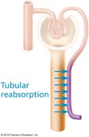
Tubular Reabsorption: Selectively returns 99% of substances from filtrate to blood.
Tubular Secretion: Selectively moves substances from blood to filtrate.
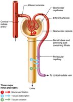
Glomerular Filtration
Mechanism of Glomerular Filtration
Glomerular filtration is a passive process driven by hydrostatic pressure. It forces fluids and solutes through the filtration membrane into the glomerular capsule, with no reabsorption occurring in the glomerulus.
The Filtration Membrane
The filtration membrane is a porous barrier between the blood and the interior of the glomerular capsule. It allows water and solutes smaller than plasma proteins to pass, while preventing the passage of cells and large proteins. The membrane consists of three layers:
Fenestrated endothelium of glomerular capillaries
Basement membrane: fused basal laminae of two other layers
Foot processes of podocytes with filtration slits and slit diaphragms
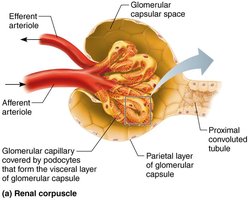
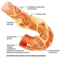
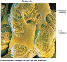
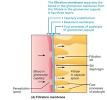
Macromolecules trapped in the membrane are engulfed by glomerular mesangial cells.
Allows passage of molecules smaller than 3 nm (e.g., water, glucose, amino acids, nitrogenous wastes).
Plasma proteins remain in the blood to maintain colloid osmotic pressure.
Presence of proteins in filtrate indicates a membrane problem.
Pressures Affecting Filtration
Filtration is influenced by both outward and inward pressures:
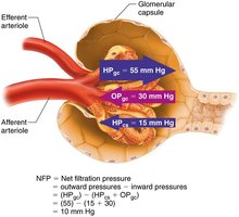
Outward pressure: Hydrostatic pressure in glomerular capillaries (HPgc) is the main force pushing water and solutes out of the blood (55 mm Hg).
Inward pressures: Hydrostatic pressure in capsular space (HPcs, 15 mm Hg) and colloid osmotic pressure in capillaries (OPgc, 30 mm Hg) oppose filtration.
Net Filtration Pressure (NFP):
Glomerular Filtration Rate (GFR)
GFR is the volume of filtrate formed per minute by the kidneys. It is a critical parameter for kidney function and is regulated by both intrinsic and extrinsic mechanisms.
Normal Rage is (120-125 ml/mim)
Hypertensive patient is going to elevate-
Regulation of Glomerular Filtration
Maintaining a constant GFR is vital for homeostasis. Regulation occurs via:
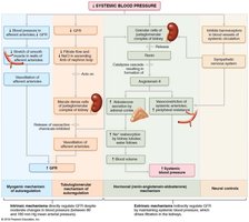
Intrinsic controls (renal autoregulation): Maintain GFR within the kidney.
Extrinsic controls: Maintain systemic blood pressure via nervous and endocrine mechanisms.
ex: Blood Pressure- Maintaining 80-180mm Hg because it can go low or high
Intrinsic Controls
Myogenic mechanism: Smooth muscle contracts when stretched, constricting afferent arterioles to protect glomeruli from high BP.
Tubuloglomerular feedback: Macula densa cells respond to NaCl concentration in filtrate, adjusting afferent arteriole diameter.
GFR Never changes constant
You can slightly constrict
Dilate afferent arteriole so the the gfr in the kidneys can dilate constant - the heart is driving the pressure not the kidneys, you open them with more systemic volume
Extrinsic Controls
Neural (sympathetic nervous system): Vasoconstriction and decreased GFR during low blood pressure.
fight or flight
Hormonal (renin-angiotensin-aldosterone mechanism): Increases blood pressure and GFR via multiple pathways.
In order to increase pressure making a pathway
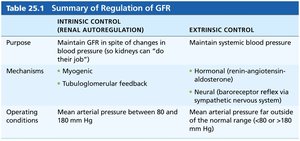
Summary Table: Regulation of GFR
Intrinsic Control (Renal Autoregulation) | Extrinsic Control |
|---|---|
Maintain GFR in spite of changes in blood pressure | Maintain systemic blood pressure |
Myogenic, Tubuloglomerular feedback | Hormonal (renin-angiotensin-aldosterone), Neural (baroreceptor reflex) |
Mean arterial pressure between 80 and 180 mm Hg | Mean arterial pressure far outside normal range (<80 or >180 mm Hg) |
Clinical Homeostatic Imbalance
Anuria: Abnormally low urinary output (<50 ml/day), may indicate low glomerular blood pressure or renal failure.
if too low you cannot create a filtrate
Causes include acute nephritis, transfusion reactions, and crush injuries.
Tubular Reabsorption
Mechanism of Tubular Reabsorption
Tubular reabsorption rapidly reclaims most of the filtrate and returns it to the blood. It is a selective transepithelial process involving both active and passive transport. Substances follow either the transcellular or paracellular route.'
Things that are carbon based are organic
Transcellular route: Solute passes through tubule cell membranes and cytosol.
Paracellular route: Solute moves between tubule cells, limited by tight junctions.
water, ions
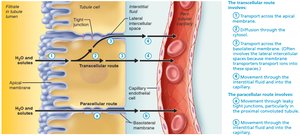 some cells are going to go through
Reabsorption of Sodium
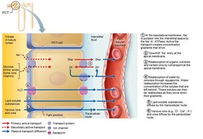
Na+ is the most abundant cation in filtrate.
Transport across basolateral membrane is via primary active transport (Na+-K+ ATPase).
Na+ is swept by bulk flow into peritubular capillaries.
Reabsorption of Nutrients, Water, and Ions
Na+ reabsorption provides energy for secondary active transport of other solutes (glucose, amino acids, ions, vitamins).
Water is reabsorbed by osmosis, aided by aquaporins.
Obligatory water reabsorption: Aquaporins always present in PCT.
Facultative water reabsorption: Aquaporins inserted in collecting ducts only if ADH is present.
Solute concentration increases as water is reabsorbed, creating gradients for passive reabsorption of solutes.
Transport Maximum (Tm)
Transcellular transport systems are specific and limited.
Tm reflects the number of carriers available for each substance.
Excess solute is excreted in urine if carriers are saturated (e.g., glucose in diabetes).
Reabsorptive Capabilities of Renal Tubules and Collecting Ducts
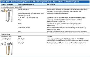
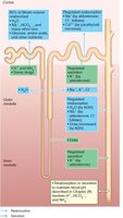
Proximal convoluted tubule (PCT): Site of most reabsorption (all nutrients, 65% Na+ and water, many ions).
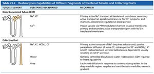
Distal convoluted tubule (DCT) and collecting duct: Reabsorption is hormonally regulated (ADH, aldosterone, atrial natriuretic peptide, parathyroid hormone).
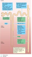
Table: Reabsorption Capabilities of Renal Tubules and Collecting Ducts
Tubule Segment | Substance Reabsorbed | Mechanism |
|---|---|---|
Proximal Convoluted Tubule (PCT) | Na+, virtually all nutrients, Cl-, K+, Mg2+, Ca2+, HCO3-, water, lipid-soluble substances | Primary and secondary active transport, passive diffusion |
Nephron Loop | Water (descending), Na+, Cl-, K+ (ascending) | Osmosis, secondary active transport |
Distal Convoluted Tubule (DCT) | Na+, Cl-, Ca2+ | Primary active transport, hormone-regulated uptake |
Collecting Duct | Na+, Cl-, HCO3-, water, urea | Primary active transport, osmosis, facilitated diffusion |
Tubular Secretion
Mechanism of Tubular Secretion
Tubular secretion is the process of moving substances from the blood into the filtrate. It occurs mainly in the PCT and involves the removal of K+, H+, NH4+, creatinine, organic acids and bases, and substances synthesized in tubule cells (e.g., HCO3-).
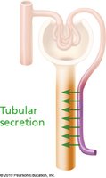
Helps control blood pH and eliminate substances not already in the filtrate.
Important for removing drugs, toxins, and excess ions.
Summary
The kidney's physiology is centered on three processes: glomerular filtration, tubular reabsorption, and tubular secretion.
Each process is tightly regulated to maintain homeostasis, blood pressure, and proper waste removal.
Hormonal and neural mechanisms play key roles in adjusting these processes according to the body's needs.
