 뒤로
Sensory Physiology: An Integrated Overview of Sensory Systems
스터디 가이드 - 스마트 노트
 자료에 맞춘 맞춤형 노트, 핵심 정의, 예시, 맥락을 확장해 제공합니다.
General Properties of Sensory Systems
Overview of Sensory Systems
The human sensory system is responsible for detecting and processing various forms of environmental stimuli. Sensory systems are divided into special senses (vision, hearing, taste, smell, equilibrium) and somatic senses (touch, temperature, pain, itch, proprioception).
Special senses are localized to specific organs.
Somatic senses are distributed throughout the body, especially in the skin and visceral organs.
Types of Sensory Receptors
Sensory receptors are specialized to detect specific types of stimuli. They can be neural or non-neural and range from free nerve endings to complex corpuscles and specialized cells.
Chemoreceptors: Respond to chemical ligands (e.g., taste, smell, blood pH).
Mechanoreceptors: Respond to physical deformation (e.g., touch, hearing, equilibrium).
Thermoreceptors: Respond to temperature changes.
Photoreceptors: Respond to light (vision).
Type of Receptor | Examples of Stimuli |
|---|---|
Chemoreceptors | Oxygen, pH, glucose |
Mechanoreceptors | Pressure, vibration, sound |
Photoreceptors | Light (photons) |
Thermoreceptors | Heat, cold |

Sensory Transduction
Sensory transduction is the process by which sensory receptors convert external stimuli into graded electrical potentials. If the graded potential reaches threshold, it triggers an action potential, leading to neurotransmitter release and signal transmission to the central nervous system (CNS).
Receptive Fields
Each sensory neuron has a receptive field, the physical area where stimuli activate the neuron. Receptive fields can overlap and may exhibit convergence, affecting sensitivity and discrimination.
Smaller receptive fields: Higher sensitivity, better two-point discrimination.
Larger receptive fields: Lower sensitivity, more convergence.
Example: The two-point discrimination test demonstrates the ability to distinguish two closely spaced stimuli as separate.
Sensory Pathways in the Brain
Most sensory pathways project to the thalamus, which relays information to specific cortical centers. Olfactory pathways are an exception, projecting directly to the olfactory cortex. Equilibrium pathways project primarily to the cerebellum.
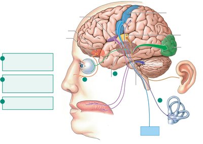
Stimulus Coding
The CNS distinguishes different stimuli by:
Modality: Which receptors and brain regions are activated.
Location: Which receptive fields are activated.
Intensity: Number and frequency of action potentials.
Duration: How long the stimulus persists; some receptors adapt quickly (phasic), others slowly (tonic).
Somatic Senses
Types and Pathways
Somatic senses include touch, proprioception, temperature, and nociception (pain and itch). Receptors are primarily located in the skin and visceral organs. Sensory information is relayed to the thalamus and then to the somatosensory cortex for integration.
Different columns in the somatosensory cortex process information from different body regions and receptor types.
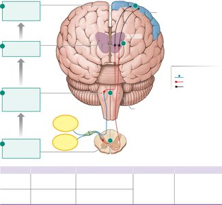
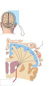
Somatosensory Cortex
The somatosensory cortex is organized such that the amount of cortical space devoted to each body part is proportional to its sensitivity. This mapping is known as the homunculus.
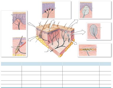
Touch Receptors
Touch receptors are mechanoreceptors that respond to various physical stimuli, such as stretch, pressure, vibration, and texture. They are found in both superficial and deep layers of the skin.
Pacinian corpuscles: Sense vibration (deep, rapid adaptation).
Meissner's corpuscles: Sense flutter and stroking (superficial, rapid adaptation).
Merkel receptors: Sense steady pressure and texture (superficial, slow adaptation).
Ruffini corpuscles: Sense skin stretch (deep, slow adaptation).
Free nerve endings: Sense temperature, noxious stimuli, and hair movement.
Thermoreceptors
Thermoreceptors are free nerve endings that terminate in the subcutaneous layers of the skin. They detect changes in temperature and are classified as cold or warm receptors. Extreme temperatures can activate nociceptors.
Nociceptors: Pain and Itch
Nociceptors are primarily chemoreceptors with free nerve endings that respond to strong, potentially damaging stimuli. They are not found in the CNS. Itch is primarily due to histamine activation of nociceptors.
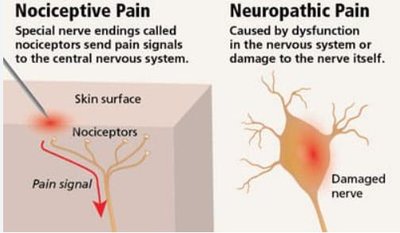
Nociceptor Pathways and Referred Pain
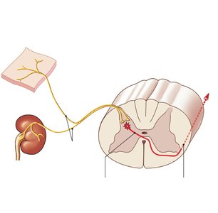
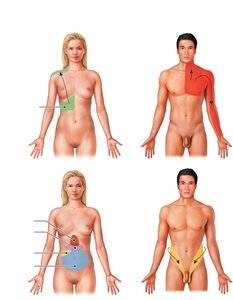
Nociceptor activation can lead to reflexive protective responses (spinal cord integration) or conscious sensation (ascending pathways to cortex). Chronic pain reflects long-term changes in the nervous system. Referred pain occurs when nociceptors from different locations converge on a single ascending tract, causing pain from internal organs to be perceived as originating from the skin.
Pain Modulation
Pain can be modulated by analgesic drugs (e.g., NSAIDs, opioids) or experimental surgical interventions. Endogenous and exogenous opioids bind to opioid receptors to decrease pain perception.
Special Senses: Olfaction (Smell)
Anatomy and Pathway
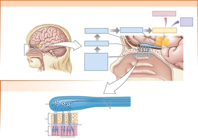
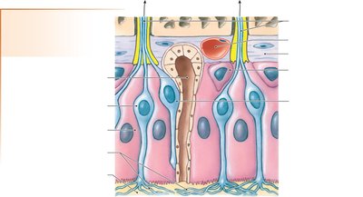
The olfactory system detects chemical stimuli (odorants) dissolved in mucus. Olfactory neurons in the nasal epithelium project to the olfactory bulb, which is an extension of the frontal lobe. Signals are relayed to the olfactory cortex and limbic system for integration.
Olfactory Transduction
Odorant molecules bind to G-protein-coupled receptors on the cilia of olfactory cells, triggering action potentials that are sent to the olfactory cortex.
Special Senses: Gustation (Taste)
Anatomy and Transduction
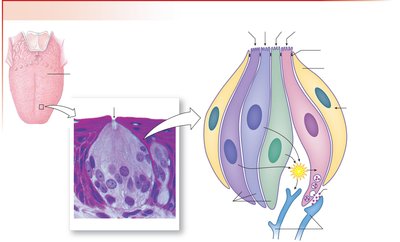
Taste buds, located on the dorsal surface of the tongue, contain gustatory (taste) cells and glial cells. Gustatory hairs protrude into the taste pore and contain chemoreceptors for molecules dissolved in saliva. Action potentials are sent to the gustatory cortex for integration.
Special Senses: Hearing and Equilibrium
Anatomy of the Ear
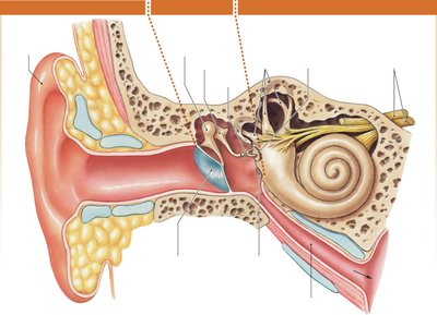
The ear is divided into three regions:
External ear: Pinna, ear canal, tympanic membrane.
Middle ear: Ossicles (malleus, incus, stapes), Eustachian tube.
Inner ear: Cochlea (hearing), vestibular apparatus and semicircular canals (equilibrium).
Cochlea and Organ of Corti
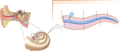
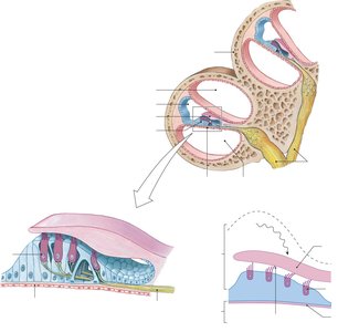
The cochlea contains three fluid-filled ducts: vestibular, cochlear, and tympanic. The cochlear duct houses the Organ of Corti, which contains hair cells (mechanoreceptors) embedded in the basilar membrane and covered by the tectorial membrane. Hair cells have stereocilia connected by protein bridges; movement opens mechanically gated K+ channels.
Sound Transduction
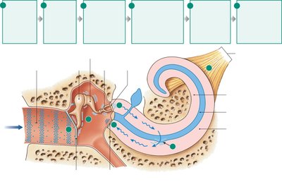
Sound waves are collected by the pinna, funneled into the ear canal, and converted to mechanical vibrations by the tympanic membrane. Vibrations are amplified by the ossicles and transferred to the oval window, creating fluid waves in the cochlea. Movement of the basilar and tectorial membranes bends hair cells, leading to action potentials sent to the auditory cortex.
Auditory Pathways
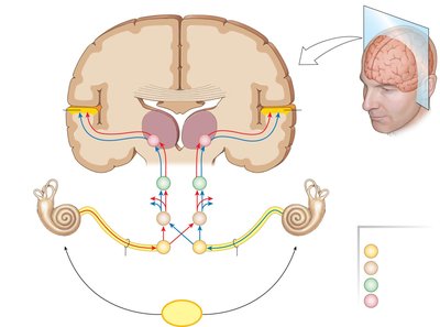
Auditory information from each ear is processed bilaterally in the brain, passing through the cochlear nuclei, superior olivary nucleus, inferior colliculus, medial geniculate body (thalamus), and finally to the auditory cortex.
Soundwave Properties and Sensory Coding
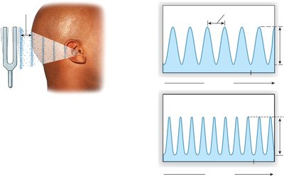
Hearing is the perception of energy carried by sound waves. The pitch of sound is determined by frequency (Hz), and loudness by amplitude (dB). The basilar membrane's variable sensitivity allows for pitch discrimination, while loudness is coded by the number and frequency of action potentials.
Types of Hearing Loss
Conductive hearing loss: Impaired transmission through external or middle ear.
Sensorineural hearing loss: Damage to inner ear structures (e.g., cochlea, hair cells).
Central hearing loss: Damage to neural pathways or auditory cortex.
Tinnitus
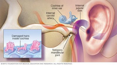
Tinnitus is the perception of ringing in the ears, often due to bent or damaged hair cells. It can be temporary or permanent.
Special Senses: Equilibrium
Maculae and Linear Acceleration
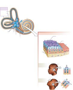
The maculae in the vestibule of the inner ear provide information about linear acceleration and head position. They consist of hair cells, the otolith membrane, and otoliths. Movement of otoliths bends hair cells, generating action potentials sent to the cerebellum.
Cristae and Rotational Acceleration
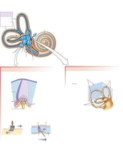
The cristae at the base of each semicircular canal sense rotational acceleration. When the head rotates, endolymph flows in the canal, pushing the cupula and bending hair cells. Both maculae and cristae send signals via the vestibulocochlear nerve to the cerebellum.
Equilibrium Pathway
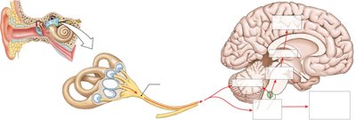
Equilibrium information is relayed from the vestibular apparatus to the vestibular nuclei of the medulla, cerebellum, thalamus, and cerebral cortex, as well as to somatic motor neurons controlling eye movements.
Special Senses: Vision
Anatomy of the Eye
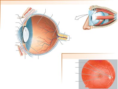
The eye consists of two chambers (anterior and vitreous), a lens for focusing light, and the iris and pupil for regulating light entry. The retina contains photoreceptors (rods and cones) embedded in the pigment epithelium, which absorbs excess light.
The Retina and Photoreceptors
The retina contains rods (for low light and peripheral vision) and cones (for high acuity and color vision, concentrated in the fovea). Photoreceptors synapse to bipolar cells, which synapse to ganglion cells, forming the optic nerve.
Visual Pigments
Photoreceptors contain visual pigments composed of retinal (a vitamin A derivative) and opsin (a structural protein). There are three types of cone pigments, each sensitive to different wavelengths of light, and rods for monochromatic vision.
Accommodation and Focusing Light
Accommodation is the process by which the ciliary body adjusts the thickness of the lens to focus light on the retina. For distant objects, the lens is flattened; for near objects, the lens becomes more rounded.
Phototransduction in the Retina
Phototransduction is the conversion of light into electrical signals by photoreceptors. In darkness, photoreceptors are depolarized and release glutamate. In light, retinal changes conformation, leading to hyperpolarization and cessation of glutamate release.
Pathway for Vision
Information from photoreceptors is relayed via bipolar and ganglion cells to the optic nerve, which crosses at the optic chiasm and projects to the visual cortex in the occipital lobe. Some collateral pathways control pupillary constriction.
Visual Defects
Myopia: Near-sightedness (inability to focus on distant objects).
Hyperopia: Far-sightedness (inability to focus on near objects).
Astigmatism: Distorted images due to misshapen cornea.
Cataracts: Protein build-up on the lens.
Glaucoma: Increased intraocular pressure damages the optic nerve.
Macular degeneration: Blood or scars block light to the retina.
