 뒤로
Skeletal Muscle Energy Sources, Fiber Types, and Performance (M2)
스터디 가이드 - 스마트 노트
 자료에 맞춘 맞춤형 노트, 핵심 정의, 예시, 맥락을 확장해 제공합니다.
Ch 10.5 - Skeletal Muscle Energy Sources
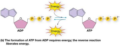
ATP: The Energy Currency of Muscle Cells
ATP (adenosine triphosphate) is the primary energy carrier in muscle cells, essential for contraction, relaxation, and maintaining ion gradients. The hydrolysis of ATP releases energy used for cellular functions.
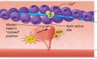
Key Point 1: ATP is required for crossbridge cycling, Ca2+ reuptake into the sarcoplasmic reticulum (SR), and the Na+/K+ pump.
Key Point 2: ATP is produced and regenerated through several metabolic pathways.
Example: The breakdown of ATP to ADP and Pi releases energy for muscle contraction.
Immediate Energy Sources: The Phosphagen System
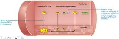
Muscle fibers utilize stored ATP and creatine phosphate for immediate energy. The phosphagen system rapidly regenerates ATP for short bursts of activity.
Key Point 1: Creatine phosphate donates a phosphate group to ADP, forming ATP.
Key Point 2: This system supports maximal muscle activity for 10-15 seconds.
Example: Sprinting or heavy lifting relies on the phosphagen system.
Cellular Metabolism: Catabolic Pathways to Make ATP
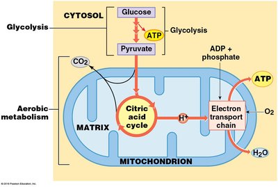
Muscle fibers generate ATP through anaerobic and aerobic metabolism. Anaerobic metabolism occurs in the cytosol without oxygen, while aerobic metabolism takes place in mitochondria and requires oxygen.
Key Point 1: Anaerobic glycolysis converts glucose to pyruvate, yielding 2 ATP per glucose.
Key Point 2: Aerobic metabolism includes the citric acid cycle and oxidative phosphorylation, producing up to 30 ATP per glucose and over 300 ATP from fatty acids.
Example: Prolonged exercise relies on aerobic metabolism for sustained ATP production.
Rapid and Sustained Sources of ATP
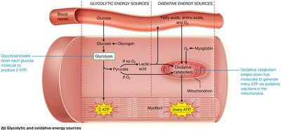
Muscle fibers use glucose, fatty acids, and occasionally amino acids as fuel for ATP production. Glycolysis provides rapid ATP, while oxidative catabolism sustains ATP production during prolonged activity.
Key Point 1: Glycolysis alone supports ~2 minutes of sustained contraction.
Key Point 2: Oxidative metabolism continues as long as oxygen and nutrients are available.
Example: Marathon running depends on oxidative metabolism.
Comparison of ATP Production Pathways
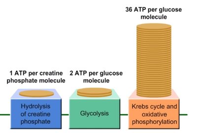
Different metabolic pathways yield varying amounts of ATP. The phosphagen system is fastest but limited, glycolysis is rapid but less efficient, and aerobic metabolism is slowest but most efficient.
Key Point 1: 1 ATP per creatine phosphate molecule (phosphagen system).
Key Point 2: 2 ATP per glucose molecule (glycolysis).
Key Point 3: ~36 ATP per glucose molecule (Krebs cycle and oxidative phosphorylation).
Example: The efficiency of ATP production increases with aerobic metabolism.
Cellular Respiration and Oxygen Consumption
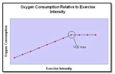
Cellular respiration is synonymous with oxidative metabolism and aerobic metabolism. Oxygen consumption (VO2) reflects the body's ability to use oxygen for ATP production.
Key Point 1: VO2 is the amount of oxygen used per minute.
Key Point 2: VO2 max is the maximum rate of oxygen use, influenced by genetics, training, and age.
Example: Elite endurance athletes have high VO2 max values.
Ch 10.6 – Classes of Skeletal Muscle Fibers
Classification of Skeletal Muscle Fibers
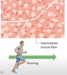
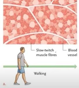
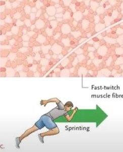
Skeletal muscle fibers are classified based on myosin ATPase activity, predominant energy source, number of mitochondria, blood supply, and myoglobin content. Most muscles contain a mix of fiber types.
Key Point 1: Type I (slow, oxidative), Type IIa (fast, oxidative-glycolytic), and Type IIx (fast, glycolytic) fibers.
Key Point 2: Fiber type distribution is influenced by genetics and muscle function.
Example: Postural muscles have more Type I fibers; sprinting muscles have more Type IIx fibers.
Myosin ATPase Activity
Myosin ATPase is an enzyme that catalyzes ATP hydrolysis during crossbridge cycling. High ATPase activity results in faster contraction speeds.
Key Point 1: Type I fibers have slow ATPase activity; Type II fibers have fast ATPase activity.
Example: Type IIx fibers are used for explosive movements.
Myoglobin Content
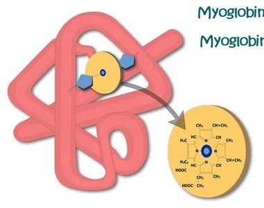
Myoglobin is a protein in muscle fibers that binds oxygen, supporting aerobic metabolism. Fibers with high myoglobin content are more resistant to fatigue.
Key Point 1: Type I fibers have high myoglobin; Type IIx fibers have low myoglobin.
Example: Myoglobin gives slow-twitch fibers a red color.
Fiber Types and Their Properties
Fiber Type | Diameter | ATPase Activity | Blood Supply | Mitochondria | Myoglobin | Fatigue Resistance |
|---|---|---|---|---|---|---|
Type I (Slow, Oxidative) | Small | Slow | High | Many | High | High |
Type IIa (Fast, Oxidative-Glycolytic) | Large | Fast | Medium | Medium | Medium | Medium |
Type IIx (Fast, Glycolytic) | Medium | Fastest | Low | Few | Low | Low |
Ch 10.8 - Skeletal Muscle Performance
Myoplasticity: Muscle Adaptation to Training
Myoplasticity refers to the ability of muscle structure to change in response to physical training. The adaptations depend on the type of training performed.
Key Point 1: Endurance training increases oxidative capacity and fatigue resistance.
Key Point 2: Resistance training increases muscle fiber diameter and strength.
Example: Combining endurance and resistance training optimizes muscle performance.
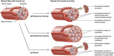
Skeletal Muscle Performance: Endurance Training
Endurance training leads to biochemical and anatomical changes that enhance aerobic metabolism and fatigue resistance.
Key Point 1: Increased mitochondria, oxidative enzymes, and blood vessel network.
Key Point 2: Enhanced oxidative capacity and resistance to fatigue.
Example: Long-distance runners exhibit these adaptations.

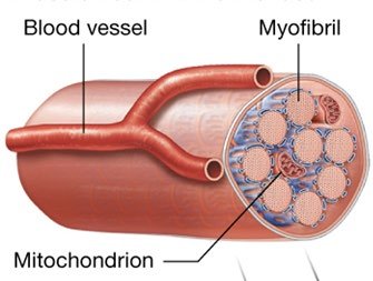
Skeletal Muscle Performance: Resistance Training
Resistance training causes anatomical changes such as increased myofibril number and muscle fiber diameter (hypertrophy), leading to greater force production.
Key Point 1: Hypertrophy decreases the proportion of mitochondrial proteins and blood supply.
Key Point 2: Excessive hypertrophy may reduce endurance.
Example: Weightlifters develop larger muscle fibers.
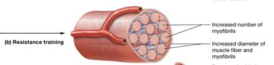
Skeletal Muscle Performance: Disuse
Disuse leads to atrophy, characterized by decreased myofibril number, fiber size, and oxidative enzymes, resulting in reduced strength and endurance.
Key Point 1: "Use it or lose it" principle applies to muscle maintenance.
Key Point 2: Atrophy is a concern for immobile individuals or those with limb loss.
Example: Prolonged bed rest causes muscle atrophy.
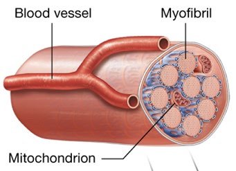

Muscle Fatigue
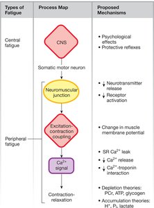
Muscle fatigue is the inability to maintain tension during activity, caused by central and peripheral factors. Accumulation of H+ lowers pH, affecting enzyme activity and Ca2+ binding.
Key Point 1: Central fatigue originates in the CNS; peripheral fatigue occurs at the neuromuscular junction or within the muscle fiber.
Key Point 2: Lowered pH impairs metabolic enzymes and Ca2+ binding to troponin.
Example: Intense exercise leads to muscle fatigue due to lactate and H+ accumulation.
 Additional info: The notes above expand on brief points with academic context, definitions, and examples to ensure completeness and clarity for exam preparation.
