 뒤로
LEC 2
스터디 가이드 - 스마트 노트
 자료에 맞춘 맞춤형 노트, 핵심 정의, 예시, 맥락을 확장해 제공합니다.
Skeletal System: Osseous Tissue & Bone Structure
Bone as a Dynamic Tissue
The skeletal system is composed of living, dynamic tissue that constantly remodels and adapts to mechanical and metabolic demands. Bone cells play distinct roles in bone formation, maintenance, and resorption.
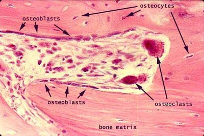
Osteoblasts: Cells responsible for bone formation; they secrete collagen and initiate mineralization.
Osteoclasts: Large, multinucleated cells that resorb bone tissue, aiding in bone remodeling and calcium release.
Osteocytes: Mature bone cells embedded in the bone matrix; they maintain bone tissue and communicate via canaliculi.
Osteoprogenitor cells: Stem cells that differentiate into osteoblasts, supporting bone growth and repair.
Bone Development & Ossification
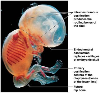
Bone formation occurs through two primary processes: intramembranous and endochondral ossification. These processes transform embryonic tissues into mature bone.
Intramembranous ossification: Produces flat bones of the skull by direct transformation of mesenchymal tissue into bone.
Endochondral ossification: Replaces hyaline cartilage with bone, forming most bones including long bones.
Primary ossification center: The initial site of bone formation in the diaphysis (shaft) of long bones.
Secondary ossification center: Forms in the epiphysis (ends) of long bones after birth.
Stages of Endochondral Ossification
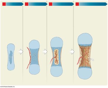
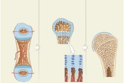
Early bones are made from hyaline cartilage.
Calcification of matrix kills chondrocytes, leaving empty lacunae.
Blood vessels develop around cartilage; perichondral cells differentiate into osteoblasts.
Perichondrium becomes periosteum; blood vessels reach the center, osteoblasts create primary ossification center.
Spongy bone forms; osteoclasts erode inside to create medullary cavity.
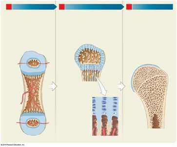
Secondary ossification centers form in epiphysis; epiphyseal plate separates epiphysis and diaphysis.
Cartilage cells in epiphyseal plate are replaced by osteoblasts; bones grow in length.
Epiphyseal plate narrows with age, eventually closing (epiphyseal line remains).
Articular cartilage persists at joint surfaces to prevent bone damage.
Bone Growth: Lengthening and Thickening
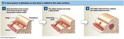
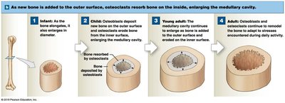
Bones grow in both length and width through distinct mechanisms. Lengthening occurs at the epiphyseal plate, while thickening (appositional growth) involves the periosteum and endosteum.
Appositional growth: Bone is added to the outer surface, increasing diameter.
Interstitial growth: Bone grows in length via the epiphyseal plate.
Periosteum: Fibrous membrane covering bone; contains osteogenic cells for appositional growth.
Endosteum: Membrane lining the inner surface; contains osteoblasts, osteoclasts, and osteoprogenitor cells.
Osteoclasts: Resorb bone from inside, enlarging the medullary cavity.
Microscopic Structure of Bone
Compact Bone
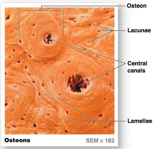
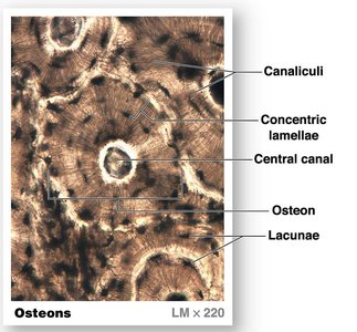
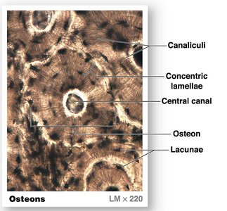
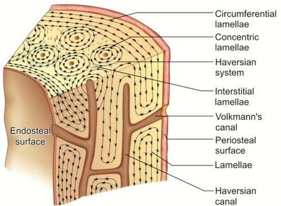
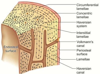
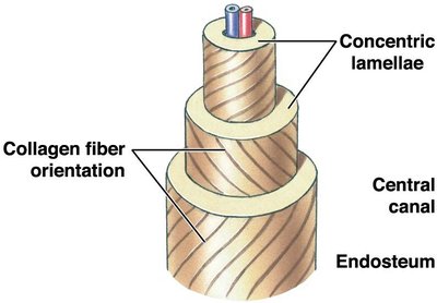
Compact bone is organized into osteons (Haversian systems), which provide strength and support. Each osteon contains concentric lamellae surrounding a central canal.
Osteon: Structural unit of compact bone.
Lamellae: Layers of bone matrix; concentric and interstitial types.
Central (Haversian) canal: Contains blood vessels and nerves.
Lacunae: Small spaces housing osteocytes.
Canaliculi: Tiny canals connecting osteocytes for nutrient and waste exchange.
Spongy Bone
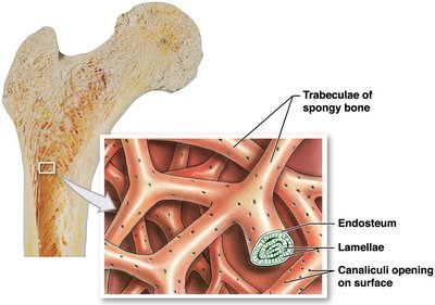
Spongy bone consists of trabeculae, which are oriented along stress lines and provide structural support with minimal weight. The spaces between trabeculae contain bone marrow.
Trabeculae: Lattice-like structures in spongy bone.
Endosteum: Lines the trabeculae and contains bone cells.
Medullary cavity: Central cavity in long bones containing marrow.
Regulation of Bone Development and Growth
Minerals, Vitamins, and Hormones
Bone growth and maintenance depend on a variety of minerals, vitamins, and hormones.
Minerals: Calcium, phosphate, magnesium, citrate, carbonate, sodium.
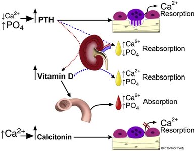
Vitamins: A and C are essential for bone health; Vitamin D3 (cholecalciferol) is required for calcium absorption.
Hormones: Parathyroid hormone (PTH), calcitonin, growth hormone, thyroxine, estrogen, and testosterone regulate bone metabolism.
Aging and the Skeletal System
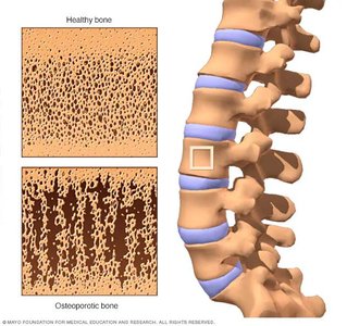
As individuals age, bone remodeling shifts, leading to decreased bone mass and increased risk of osteoporosis.
Osteopenia: Reduced bone mass due to decreased ossification.
Osteoporosis: Severe bone loss resulting in porous, fragile bones; more common in postmenopausal women due to decreased estrogen.
Classification of Skeletal Elements
Bone Types by Shape
Bones are classified into seven broad categories based on their shape and function.
Sutural bones: Small, irregular bones found between cranial sutures.
Irregular bones: Complex shapes (e.g., vertebrae).
Short bones: Cube-shaped (e.g., wrist bones).
Pneumatized bones: Bones with air-filled spaces (e.g., ethmoid).
Flat bones: Thin, broad surfaces (e.g., skull, ribs).
Long bones: Longer than wide (e.g., femur, humerus).
Sesamoid bones: Small, round bones embedded in tendons (e.g., patella).
Summary Table: Bone Cells and Functions
Cell Type | Function |
|---|---|
Osteoblast | Bone formation |
Osteoclast | Bone resorption |
Osteocyte | Maintenance of bone matrix |
Osteoprogenitor | Stem cell for bone repair |
Summary Table: Bone Growth Mechanisms
Growth Type | Location | Result |
|---|---|---|
Appositional | Periosteum | Increased diameter |
Interstitial | Epiphyseal plate | Increased length |
Key Equations
Calcium and phosphate regulation in bone:
Conclusion
The skeletal system is a complex, dynamic structure essential for support, protection, movement, and mineral homeostasis. Understanding bone cells, growth mechanisms, microscopic structure, and regulatory factors is fundamental for Anatomy & Physiology students.
