 뒤로
Study Guide: Blood – Structure, Function, and Physiology
스터디 가이드 - 스마트 노트
 자료에 맞춘 맞춤형 노트, 핵심 정의, 예시, 맥락을 확장해 제공합니다.
The Blood: Structure and Function
Overview of Blood
Blood is the only liquid connective tissue in the human body, essential for transport, defense, and homeostasis. It consists of formed elements (cells and cell fragments) and plasma (the liquid matrix).
Viscosity: Blood is five times more viscous than water due to its solid components.
Volume: Average adult blood volume is 4-6 liters.
Functions: Transport of gases, nutrients, wastes, hormones; stabilization of pH and electrolytes; restriction of fluid loss; defense against toxins/pathogens; stabilization of body temperature.
Components of Blood
Blood is composed of formed elements and plasma. Formed elements include cells (erythrocytes, leukocytes) and cell fragments (platelets). Plasma is a mixture of water, electrolytes, proteins, nutrients, wastes, hormones, and gases.
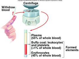
Formed Elements: Slightly less than half the blood volume.
Plasma: Represents about 55% of blood volume; 92% water.
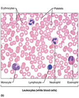
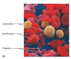
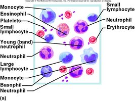
Blood Composition Table
Component | Percentage | Main Function |
|---|---|---|
Plasma | ~55% | Transport, osmotic balance |
Erythrocytes | ~45% | Oxygen and CO2 transport |
Leukocytes | <1% | Defense |
Platelets | <1% | Clotting |
Plasma: Composition and Functions
Plasma Proteins
Plasma contains several proteins, each with distinct functions:
Albumins (≈60%): Most abundant; contribute to viscosity, osmolarity, and influence blood pressure and fluid balance.
Globulins (≈35%): Immune system defenses; includes alpha, beta, and gamma globulins.
Fibrinogen (≈4%): Precursor to fibrin, essential for blood clotting.
Regulatory proteins: <1%; includes hormones and enzymes.
Most plasma proteins are produced by the liver, except immunoglobulins (by immune cells) and regulatory proteins.
Nonprotein Components
Amino acids: From dietary protein or tissue breakdown.
Nitrogenous wastes: Urea, removed by kidneys.
Nutrients: Glucose, vitamins, fats, minerals.
Blood gases: O2 and CO2.
Electrolytes: Na+ (90% of plasma cations), Cl-, HCO3-.
Formed Elements of Blood
Hematocrit
Hematocrit is the percentage of whole blood occupied by cellular components, mainly red blood cells. The ratio of RBCs to WBCs is approximately 1000:1.
Red Blood Cells (Erythrocytes)
Structure and Function
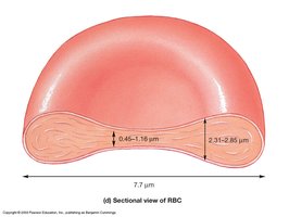
Red blood cells are the most abundant formed elements, specialized for oxygen and carbon dioxide transport. Their biconcave disc shape increases surface-to-volume ratio, facilitating gas exchange.
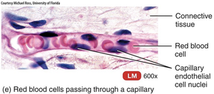
Shape: Biconcave disc, flexible for capillary passage.
Composition: 33% cytoplasm is hemoglobin.
Enzyme: Contains carbonic anhydrase, catalyzing formation of carbonic acid from CO2 and water.
Hemoglobin
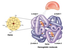
Hemoglobin is a globular protein with four globin subunits, each associated with a heme molecule. Each heme contains an Fe2+ ion that binds O2 reversibly.
Adult Hemoglobin: Two alpha chains, two beta chains.
Oxygen Binding: Each chain holds one iron-containing heme group.
RBC Count and Hemoglobin Concentration
RBC Count: Men: 4.6-6.2 million/μL; Women: 4.2-5.4 million/μL.
Hemoglobin Concentration: Men: 13-18 g/dL; Women: 12-16 g/dL.
Factors: Androgens stimulate RBC production; menstrual losses lower values in women.
Hemopoiesis: Blood Cell Formation
Overview
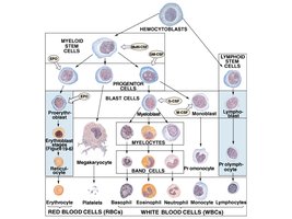
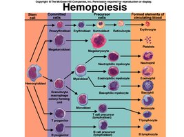
Hemopoiesis (hematopoiesis) is the formation of blood's formed elements, occurring in red bone marrow from hemocytoblasts. These stem cells differentiate into all blood cell types.
Erythropoiesis
Erythropoiesis is the production of RBCs, occurring in myeloid tissue. It produces about 2.5 million RBCs/sec from hemocytoblasts.
Stages: Proerythroblast (first committed cell), erythroblast (hemoglobin synthesis), normoblast (nucleus discarded), reticulocyte (enters bloodstream).
Development: Takes 3-5 days; blood loss increases reticulocyte count.
Regulation of Erythropoiesis
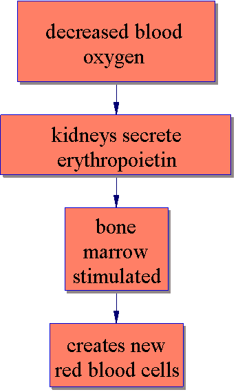
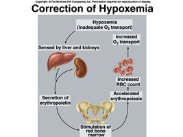
Erythropoiesis is regulated by erythropoietin (EPO) from the kidneys in response to hypoxemia (low blood oxygen).
Stimulus: Low O2, increased exercise, hemorrhage.
Negative Feedback: Drop in RBC count triggers EPO production, stimulating bone marrow.
Iron Intake and Erythropoiesis
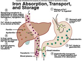
Dietary iron is essential for hemoglobin synthesis. Iron is absorbed, transported, and stored in various forms.
Forms: Ferric (Fe3+) and ferrous (Fe2+).
Transport: Gastroferritin binds Fe2+ for absorption; transferrin transports iron in blood; ferritin stores iron in liver.
RBC Lifespan and Breakdown
Lifespan
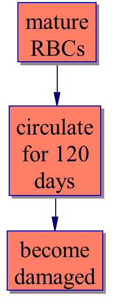
RBCs circulate for about 120 days before becoming damaged due to their inability to synthesize new membrane components.
Breakdown and Disposal
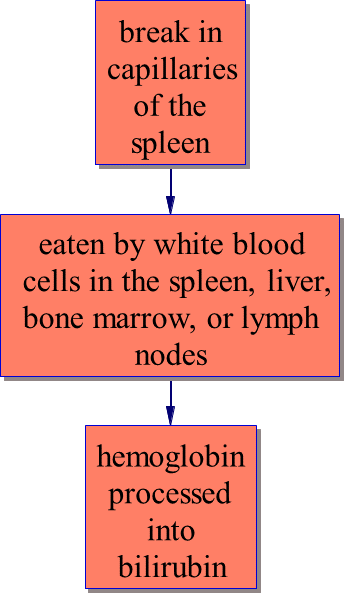
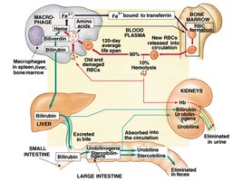
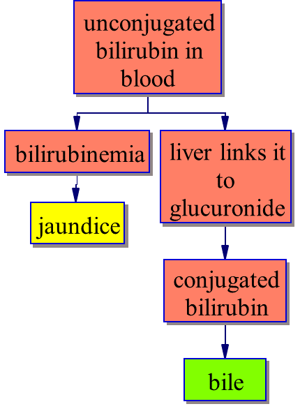
Most RBCs break in the spleen, where macrophages process damaged cells. Heme is converted to biliverdin and then bilirubin, which is excreted in bile.
Hemolysis: Breakdown occurs in narrow channels of the spleen.
Macrophages: Digest membrane, separate heme from globin, hydrolyze globin, remove iron, convert heme to biliverdin/bilirubin.
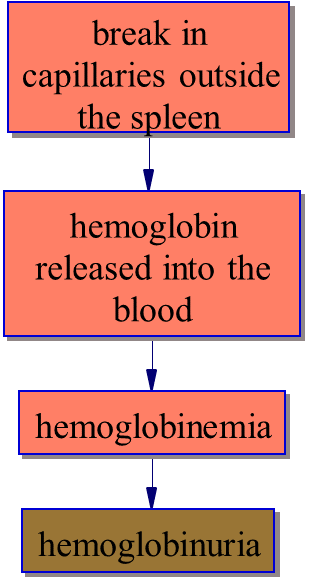
RBC Destruction Outside the Spleen
When RBCs are destroyed outside the spleen, hemoglobin is released into the blood, causing hemoglobinemia and hemoglobinuria.
Blood Types and Clinical Applications
ABO Blood Types
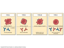
Blood type is determined by the presence or absence of antigens (agglutinogens) A and B on RBC membranes. Antibodies (agglutinins) appear in plasma.
Type A: Surface antigen A, anti-B antibodies.
Type B: Surface antigen B, anti-A antibodies.
Type AB: Surface antigens A and B, no antibodies.
Type O: No antigens, anti-A and anti-B antibodies.
Rh Factor
The Rh factor is another antigen on RBCs. Blood is Rh+ if agglutinogens are present. Rh incompatibility can cause hemolytic disease of the newborn.
White Blood Cells (Leukocytes)
Structure and Function
Leukocytes defend the body against pathogens and remove toxins, wastes, and abnormal cells. They are grouped into two classes:
Granular leukocytes: Neutrophils, eosinophils, basophils.
Agranular leukocytes: Lymphocytes, monocytes.
Leukopoiesis
Leukopoiesis is the formation of white blood cells, regulated by colony-stimulating factors (CSFs) released in response to infection.
Platelets (Thrombocytes)
Structure and Function
Platelets are membrane-enclosed fragments of megakaryocytes, essential for clotting and vessel repair.
Functions: Secrete clotting factors, form temporary plugs, dissolve clots, phagocytize bacteria, attract WBCs.
Thrombopoiesis
Thrombopoiesis is the production of platelets, stimulated by thrombopoietin from the liver or kidney.
Hemostasis: Blood Clotting
Mechanisms
Hemostasis is effective at closing breaks in small vessels and involves three mechanisms:
Vascular Spasm: Prompt constriction of a broken vessel.
Platelet Plug Formation: Platelets stick to damaged vessel, release substances to attract more platelets.
Coagulation: Final phase; intrinsic and extrinsic pathways converge to form fibrin network.
Summary Table: Blood Cell Types
Cell Type | Structure | Main Function |
|---|---|---|
Erythrocyte | Biconcave, no nucleus | O2 and CO2 transport |
Leukocyte | Varied, nucleus present | Defense, immunity |
Platelet | Fragment, no nucleus | Clotting, vessel repair |
Example: A transfusion reaction occurs if incompatible blood types are mixed, leading to agglutination and hemolysis.
Additional info: The notes include expanded explanations, definitions, and context for all major blood components and processes, suitable for exam preparation in Anatomy & Physiology.
