 뒤로
Ch. 12 Part 1
스터디 가이드 - 스마트 노트
 자료에 맞춘 맞춤형 노트, 핵심 정의, 예시, 맥락을 확장해 제공합니다.
The Central Nervous System: Cerebrum and Cerebellum
Brain Regions and Organization
The adult human brain is organized into four primary regions, each with distinct anatomical and functional characteristics:
Cerebral Hemispheres
Diencephalon
Brain Stem (composed of the midbrain, pons, and medulla oblongata)
Cerebellum
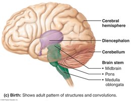
These regions are interconnected and work together to regulate sensory input, motor output, and higher cognitive functions.
Gray Matter and White Matter in the CNS
The central nervous system (CNS) is characterized by a specific pattern of gray and white matter distribution:
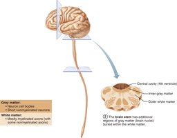
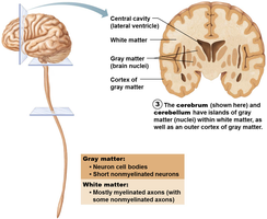
Gray Matter: Composed of neuron cell bodies and short, nonmyelinated neurons.
White Matter: Consists mainly of myelinated axons, with some nonmyelinated axons.
Basic Pattern: Central cavity surrounded by gray matter, with white matter external to gray matter.
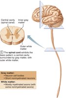
As the CNS ascends from the spinal cord to the brain stem and cerebrum, the pattern changes, with additional gray matter nuclei appearing within the white matter.
Ventricles of the Brain
The brain contains fluid-filled chambers called ventricles that are continuous with each other and the central canal of the spinal cord. These ventricles are filled with cerebrospinal fluid (CSF) and lined by ependymal cells.
Lateral Ventricles: Paired, C-shaped chambers deep in each hemisphere, separated by the septum pellucidum.
Third Ventricle: Located in the diencephalon, connected to lateral ventricles via the interventricular foramen.
Fourth Ventricle: Located in the hindbrain, connected to the third ventricle via the cerebral aqueduct; continuous with the central canal of the spinal cord.
Apertures: Three openings connect the fourth ventricle to the subarachnoid space (paired lateral apertures and a median aperture).
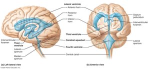
Cerebral Hemispheres
Surface Markings and Lobes
The cerebral hemispheres form the superior part of the brain and account for 83% of its mass. The surface is marked by:
Gyri: Ridges
Sulci: Shallow grooves
Fissures: Deep grooves (e.g., longitudinal fissure separates hemispheres; transverse cerebral fissure separates cerebrum and cerebellum)
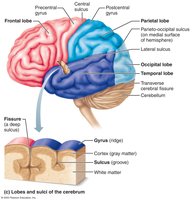
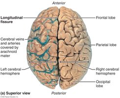
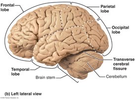
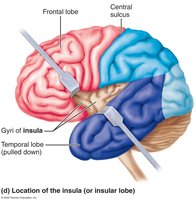
Each hemisphere is divided into five lobes: frontal, parietal, temporal, occipital, and insula (the insular lobe is buried under portions of the other lobes).
Major Sulci and Regions
Central Sulcus: Separates precentral gyrus (frontal lobe) and postcentral gyrus (parietal lobe).
Parieto-occipital Sulcus: Separates occipital and parietal lobes.
Lateral Sulcus: Outlines temporal lobes.
Each hemisphere contains three basic regions:
Cerebral Cortex: Superficial layer of gray matter
White Matter: Internal
Basal Nuclei: Deep within white matter
Cerebral Cortex
Structure and Function
The cerebral cortex is the "executive suite" of the brain, responsible for conscious mind functions such as awareness, sensory perception, voluntary motor initiation, communication, memory storage, and understanding. It is a thin (2–4 mm) layer of gray matter composed of neuron cell bodies, dendrites, glial cells, and blood vessels.
Functional imaging (PET and MRI) reveals that specific motor and sensory functions are localized in discrete cortical areas called domains, while higher functions are distributed across multiple areas.
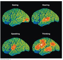
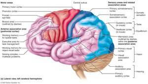
Functional Areas of the Cortex
Motor Areas: Control voluntary movement (located in frontal lobe)
Sensory Areas: Conscious awareness of sensation
Association Areas: Integrate diverse information
Each hemisphere is concerned with the contralateral (opposite) side of the body. Lateralization of function can occur in only one hemisphere, and conscious behavior involves the entire cortex.
Motor Areas
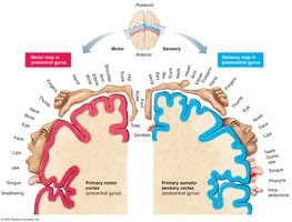
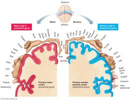
Primary Motor Cortex: Located in the precentral gyrus; contains pyramidal cells for conscious control of precise, skilled movements. Somatotopy maps all muscles to specific cortex areas.
Premotor Cortex: Plans movements, controls learned and patterned motor skills, coordinates actions, and integrates sensory feedback.
Broca’s Area: Motor speech area, usually in the left hemisphere; directs muscles of speech production.
Frontal Eye Field: Controls voluntary eye movements.
Clinical Note: Motor Cortex Damage
Damage to the primary motor cortex (e.g., stroke) results in paralysis of muscles controlled by those areas, affecting the opposite side of the body.
Muscle strength is not impaired, but voluntary control is lost. Other neurons can be reprogrammed with practice.
Sensory Areas
Primary Somatosensory Cortex: Located in postcentral gyri; receives sensory information from skin and proprioceptors. Capable of spatial discrimination.
Somatosensory Association Cortex: Integrates sensory input for object understanding (size, texture, relationship).
Visual Areas: Primary visual cortex (occipital lobe); visual association area interprets visual stimuli.
Auditory Areas: Primary auditory cortex (temporal lobe); association area stores sound memories.
Vestibular Cortex: Responsible for balance awareness.
Olfactory Cortex: Conscious awareness of odors.
Gustatory Cortex: Perception of taste.
Visceral Sensory Area: Perception of visceral sensations (e.g., upset stomach).
Clinical Note: Visual Cortex Damage
Damage to the primary visual cortex results in functional blindness.
Damage to the visual association area allows sight but impairs comprehension of visual stimuli.
Multimodal Association Areas
Anterior Association Area (Prefrontal Cortex): Involved in intellect, cognition, recall, personality, judgment, reasoning, and planning.
Posterior Association Area: Recognizes patterns and faces, localizes in space, and understands language (Wernicke’s area).
Limbic Association Area: Provides emotional impact and helps establish memories.
Clinical Note: Association Area Lesions
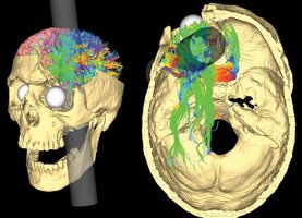
Lesions in the anterior association area may cause mental and personality disorders.
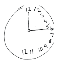
Lesions in the posterior association area may cause hemineglect (lack of awareness of one side of the body).
Lateralization of Cortical Functioning
Lateralization: Division of labor between hemispheres; hemispheres are not identical.
Cerebral Dominance: Refers to the hemisphere dominant for language (90% left-sided dominance, usually right-handedness).
Left Hemisphere: Controls language, math, and logic.
Right Hemisphere: Controls visual-spatial skills, intuition, emotion, artistic and musical skills.
Hemispheres communicate via fiber tracts and functional integration.
Cerebral White Matter
Structure and Fiber Types
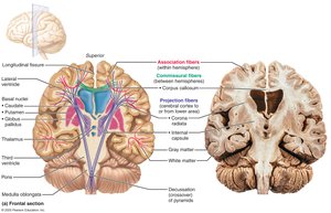
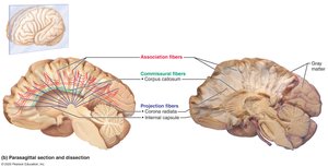
Cerebral white matter is responsible for communication between cerebral areas and between the cortex and lower CNS. It consists of myelinated fibers bundled into large tracts, classified by direction:
Association Fibers: Connect different parts of the same hemisphere.
Commissural Fibers: Connect gray matter of two hemispheres.
Projection Fibers: Connect hemispheres with lower brain or spinal cord.
Internal Capsule: Compact band of projection fibers passing between thalamus and basal nuclei.
Corona Radiata: Projection fibers radiating through cerebral white matter to cortex.
Basal Nuclei (Ganglia)
Structure and Function
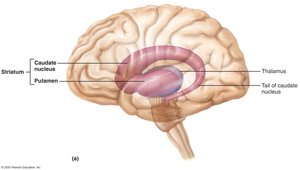
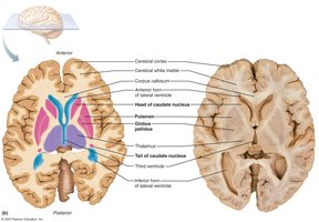
The basal nuclei are deep gray matter structures in each hemisphere, including:
Caudate Nucleus
Putamen
Globus Pallidus
Caudate nucleus + putamen = striatum
Functions:
Influence muscle movements
Play a role in cognition and emotion
Regulate intensity of slow or stereotyped movements
Filter out incorrect/inappropriate responses
Inhibit antagonistic/unnecessary movements
Disorders: Parkinson’s disease and Huntington’s disease
Cerebellum
Structure and Function
The cerebellum constitutes 11% of brain mass and is located dorsal to the pons and medulla. It processes input from the cortex, brain stem, and sensory receptors to provide precise, coordinated movements of skeletal muscles and plays a major role in balance.
Cerebellar Anatomy
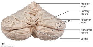
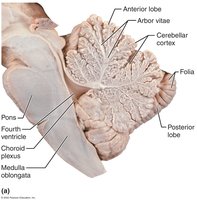
Cerebellar hemispheres are connected by the vermis.
Folia: Transversely oriented gyri.
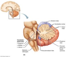
Each hemisphere has three lobes: anterior, posterior, and flocculonodular.
Contains a thin cortex of gray matter with a distinctive treelike pattern of white matter called arbor vitae.
Cerebellar Peduncles
All fibers in the cerebellum are ipsilateral (from and to the same side of the body).
Three paired fiber tracts connect the cerebellum to the brain stem:
Superior Cerebellar Peduncles: Connect cerebellum to midbrain.
Middle Cerebellar Peduncles: Connect pons to cerebellum.
Inferior Cerebellar Peduncles: Connect medulla to cerebellum.
Cerebellar Processing
Cerebellum fine-tunes motor activity by receiving impulses from the cerebral cortex regarding voluntary muscle contraction.
Receives signals from proprioceptors, visual, and equilibrium pathways to continuously inform the cerebellum of body position and momentum.
Cerebellar cortex calculates the best way to coordinate muscle contraction and sends a "blueprint" of movement to the cerebral motor cortex and brain stem nuclei.
Summary Table: Major Brain Regions and Functions
Region | Main Function |
|---|---|
Cerebral Hemispheres | Conscious thought, voluntary movement, sensory perception |
Diencephalon | Relay and processing centers for sensory information |
Brain Stem | Basic life functions, pathway for information |
Cerebellum | Coordination of movement, balance |
