 뒤로
Study Guide: The Respiratory System (Anatomy & Physiology)
스터디 가이드 - 스마트 노트
 자료에 맞춘 맞춤형 노트, 핵심 정의, 예시, 맥락을 확장해 제공합니다.
The Respiratory System
Overview and Importance
The respiratory system is essential for gas exchange, supplying oxygen to the body and removing carbon dioxide. It works closely with the circulatory system and also plays roles in olfaction and speech.
Key Functions: Gas exchange, olfaction, speech production
Interaction: Closely coupled with circulatory system for transport of gases
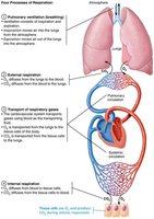
Anatomy of the Respiratory System
The respiratory system is divided into upper and lower regions, each with distinct structures and functions.
Upper Respiratory Tract: Nose, nasal cavity, paranasal sinuses, pharynx
Lower Respiratory Tract: Larynx, trachea, bronchi, lungs, alveoli
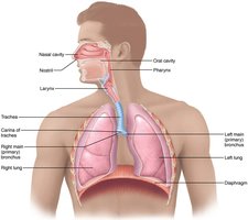
Upper Respiratory System
The Nose and Paranasal Sinuses
The nose is the only external part of the respiratory system and serves several important functions.
Functions: Provides airway, moistens and warms air, filters and cleans air, serves as resonating chamber for speech, houses olfactory receptors
Paranasal Sinuses: Located in frontal, sphenoid, ethmoid, and maxillary bones; lighten skull, secrete mucus, help warm and moisten air
Nasal Conchae: Filter, heat, and moisten air during inhalation; reclaim heat and moisture during exhalation
The Pharynx
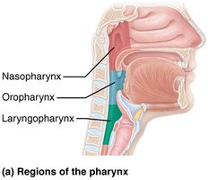
The pharynx is a muscular tube connecting the nasal cavity and mouth to the larynx and esophagus. It is divided into three regions: nasopharynx, oropharynx, and laryngopharynx.
Structure: Funnel-shaped, composed of skeletal muscle
Function: Passageway for air and food
Lower Respiratory System
Zones of the Lower Respiratory System
The lower respiratory system is divided into two functional zones:
Conducting Zone: Transports air to and from gas exchange sites; includes larynx, trachea, bronchi, and larger bronchioles; cleanses, warms, and humidifies air
Respiratory Zone: Site of gas exchange; includes respiratory bronchioles, alveolar ducts, and alveoli
The Larynx
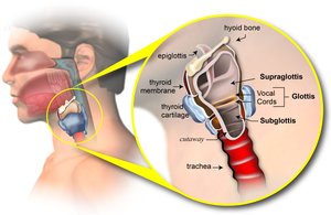
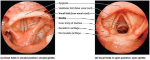
The larynx, or voice box, is involved in airway maintenance, routing air and food, and voice production.
Structure: Attaches to hyoid bone, extends from 3rd to 6th cervical vertebra
Functions: Provides patent airway, routes air and food, houses vocal folds for sound production
Epiglottis: Elastic cartilage that covers laryngeal inlet during swallowing
The Trachea
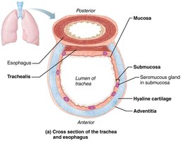
The trachea, or windpipe, is a flexible tube that conducts air from the larynx to the bronchi.
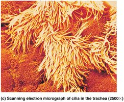
Structure: About 4 inches long, composed of three layers: mucosa (ciliated pseudostratified epithelium with goblet cells), submucosa, and hyaline cartilage
Trachealis: Smooth muscle fibers that contract during coughing
Carina: Last tracheal cartilage, highly sensitive, triggers coughing if foreign objects contact it
The Bronchi and Subdivisions
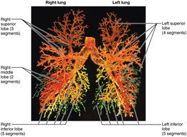
The bronchi undergo extensive branching, forming the bronchial tree. The conducting zone gives rise to the respiratory zone.
Branching: 23 orders of branching; conducting zone structures lead to respiratory zone structures
Respiratory Zone: Terminal bronchioles feed into respiratory bronchioles, alveolar ducts, and alveolar sacs
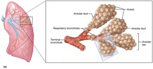
Alveoli and the Respiratory Membrane
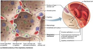
Alveoli are the primary sites of gas exchange. The respiratory membrane is a thin barrier allowing diffusion of gases between alveoli and capillaries.
Structure: Alveolar walls consist of type I (squamous) and type II (cuboidal) alveolar cells; type II cells secrete surfactant and antimicrobial proteins
Respiratory Membrane: Composed of alveolar and capillary walls with fused basement membranes; very thin (~0.5 μm)
Function: Allows gas exchange by simple diffusion
Lungs and Pleurae
Gross Anatomy of the Lungs
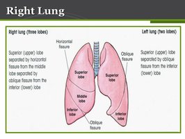
The lungs are divided into lobes and bronchopulmonary segments, each served by its own artery, vein, and bronchus.
Lobes: Right lung has 3 lobes; left lung has 2 lobes
Bronchopulmonary Segments: 10 on right, 8–10 on left; separated by connective tissue septa
Lobules: Smallest subdivisions visible to naked eye; hexagonal segments served by bronchioles
Composition: Mostly alveoli, with elastic connective tissue (stroma) making lungs elastic and spongy
The Pleurae
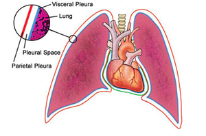
The pleurae are thin, double-layered serosal membranes that divide the thoracic cavity and provide lubrication for lung movement.
Structure: Visceral pleura covers lungs; parietal pleura lines thoracic cavity
Pleural Fluid: Fills pleural cavity, provides lubrication and surface tension for expansion and recoil of lungs
Clinical Homeostatic Imbalances
Several conditions can affect the respiratory system, including rhinitis, sinusitis, laryngitis, pleurisy, and pleural effusion.
Rhinitis: Inflammation of nasal mucosa, can spread to sinuses and tear ducts
Laryngitis: Inflammation of vocal folds, often caused by infection or overuse
Pleurisy: Inflammation of pleurae, causing pain and friction during breathing
Pleural Effusion: Accumulation of fluid in pleural cavity, hindering breathing
Summary Table: Major Structures and Functions of the Respiratory System
Structure | Main Function |
|---|---|
Nose | Airway, warms/moistens/filters air, olfaction |
Pharynx | Passageway for air and food |
Larynx | Voice production, airway maintenance |
Trachea | Conducts air, filters particles |
Bronchi | Air distribution |
Alveoli | Gas exchange |
Lungs | Contain alveoli, gas exchange |
Pleurae | Lubrication, compartmentalization |
Key Equations
Gas exchange across the respiratory membrane is governed by simple diffusion:
Fick's Law of Diffusion:
Where J is the rate of diffusion, D is the diffusion coefficient, P_1 and P_2 are partial pressures, and d is membrane thickness.
Additional info: The notes have been expanded with academic context and definitions for clarity and completeness.
