 뒤로
The Endocrine System: Structure, Function, and Regulation
스터디 가이드 - 스마트 노트
 자료에 맞춘 맞춤형 노트, 핵심 정의, 예시, 맥락을 확장해 제공합니다.
The Endocrine System
Overview and Functions
The endocrine system works alongside the nervous system to coordinate and integrate the activity of body cells. It influences metabolic activities through hormones transported in the blood. Endocrine responses are slower but longer-lasting compared to nervous system responses.
Hormones: Chemical messengers released into the blood, affecting distant target cells.
Major functions: Reproduction, growth and development, maintenance of electrolyte, water, and nutrient balance, regulation of cellular metabolism and energy balance, and mobilization of body defenses.
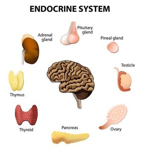
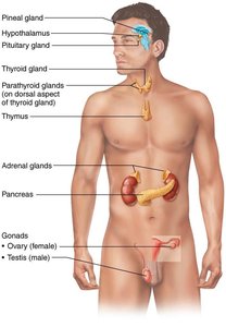
Comparison of Nervous and Endocrine Systems
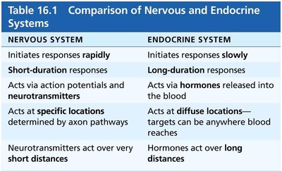
The nervous and endocrine systems both regulate body functions, but differ in their mechanisms and speed of action.
Nervous System | Endocrine System |
|---|---|
Initiates responses rapidly | Initiates responses slowly |
Short-duration responses | Long-duration responses |
Acts via action potentials and neurotransmitters | Acts via hormones released into the blood |
Acts at specific locations | Acts at diffuse locations |
Neurotransmitters act over very short distances | Hormones act over long distances |
Endocrine Glands and Hormones
Types of Glands
Exocrine glands: Produce nonhormonal substances (e.g., sweat, saliva) and have ducts.
Endocrine glands: Produce hormones, lack ducts, and release hormones into surrounding tissue fluid.
Major endocrine glands include the pituitary, thyroid, parathyroid, adrenal, and pineal glands. The hypothalamus is a neuroendocrine organ. Some organs, such as the pancreas, gonads, and placenta, have both endocrine and exocrine functions.
Chemical Messengers
Hormones: Long-distance chemical signals traveling in blood or lymph.
Autocrines: Affect the same cells that secrete them.
Paracrines: Affect nearby cells other than those that secrete them.
Autocrines and paracrines are local messengers and not considered part of the endocrine system.
Classes of Hormones
Amino acid–based hormones: Includes derivatives, peptides, and proteins; usually water-soluble and cannot cross cell membranes.
Steroid hormones: Synthesized from cholesterol; lipid-soluble and can cross cell membranes; only gonadal and adrenocortical hormones.
Eicosanoids: Sometimes considered a third class, but mostly classified as paracrine.
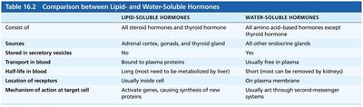
Lipid-Soluble Hormones | Water-Soluble Hormones |
|---|---|
All steroid hormones and thyroid hormone | All amino acid-based hormones except thyroid hormone |
Adrenal cortex, gonads, thyroid gland | Other endocrine glands |
Bound to plasma proteins | Usually free in plasma |
Long half-life | Short half-life |
Usually inside cell | On plasma membrane |
Activate genes, causing synthesis of new proteins | Act through second-messenger systems |
Hormone Action and Regulation
Hormone Action on Target Cells
Hormones affect only cells with specific receptors (target cells). They alter target cell activity by:
Changing membrane permeability or potential
Stimulating synthesis of enzymes or proteins
Activating or deactivating enzymes
Inducing secretory activity
Stimulating mitosis
Mechanisms of Hormone Action
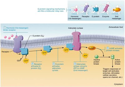
Water-soluble hormones: Act on plasma membrane receptors via G protein second messengers (e.g., cyclic AMP, PIP2-calcium).
Lipid-soluble hormones: Act on intracellular receptors that directly activate genes.
Endocrine Gland Stimuli
Endocrine glands are stimulated to release hormones by:
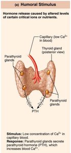
Humoral stimuli: Changes in blood levels of ions or nutrients.
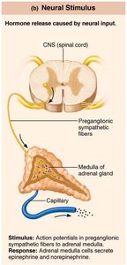
Neural stimuli: Nerve fibers stimulate hormone release.
Hormonal stimuli: Hormones stimulate other endocrine organs to release hormones.
Hormone Release Regulation
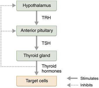
Blood levels of hormones are controlled by negative feedback systems. Hormone release is triggered by endocrine gland stimuli and nervous system modulation.
Target Cell Specificity
Target cell activation depends on:
Blood levels of hormone
Number of receptors on/in target cell
Affinity of binding between receptor and hormone
Up-regulation increases receptor number in response to low hormone levels; down-regulation decreases receptor number in response to high hormone levels.
Hormone Half-Life, Onset, and Duration
Half-life: Time required for hormone level in blood to decrease by half.
Water-soluble hormones have shorter half-lives than lipid-soluble hormones.
Onset and duration of hormone activity vary depending on hormone type.
Hormone Interactions at Target Cells
Permissiveness: One hormone cannot exert its full effects without another hormone present.
Synergism: More than one hormone produces the same effect, causing amplification.
Antagonism: One or more hormones oppose the action of another hormone.
Hypothalamus and Pituitary Gland
Structure and Function
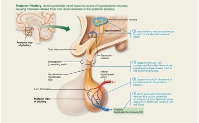
The hypothalamus is connected to the pituitary gland via the infundibulum. The pituitary gland has two lobes:
Posterior pituitary: Neural tissue; stores and releases neurohormones from the hypothalamus.
Anterior pituitary: Glandular tissue; manufactures and releases hormones.
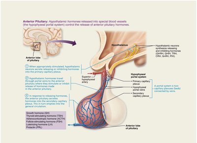
Posterior Pituitary Hormones
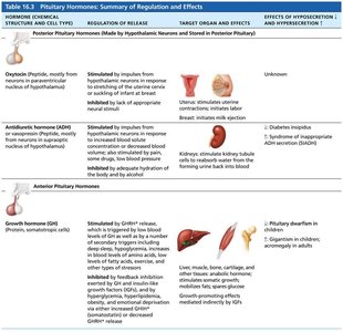
Oxytocin: Stimulates uterine contractions and milk ejection; acts via positive feedback.
Antidiuretic hormone (ADH): Regulates water balance; targets kidney tubules to reabsorb water.
Anterior Pituitary Hormones
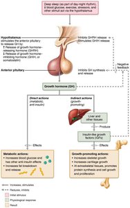
Growth hormone (GH): Stimulates growth and metabolic functions.
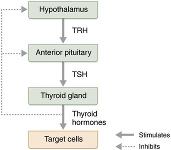
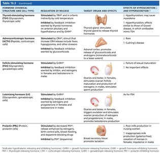
Thyroid-stimulating hormone (TSH): Stimulates thyroid gland.
Adrenocorticotropic hormone (ACTH): Stimulates adrenal cortex.
Follicle-stimulating hormone (FSH): Stimulates gamete production.
Luteinizing hormone (LH): Stimulates production of gonadal hormones.
Prolactin (PRL): Stimulates milk production.
Thyroid and Parathyroid Glands
Thyroid Gland Structure
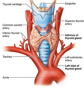
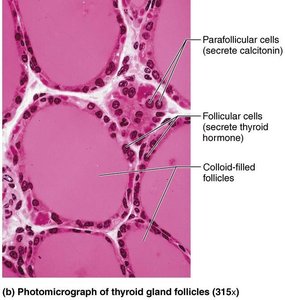
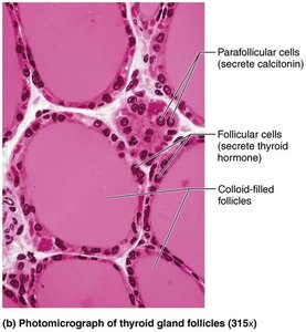
The thyroid gland is a butterfly-shaped gland located in the anterior neck, just inferior to the larynx. It consists of follicles, colloid, and parafollicular cells.
Thyroid Hormone (TH)
T4 (thyroxine): Major form; two tyrosine molecules with four iodine atoms.
T3 (triiodothyronine): Two tyrosines with three iodine atoms; more active than T4.
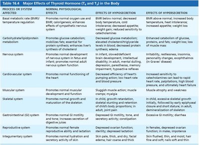
TH increases basal metabolic rate, regulates tissue growth, and maintains blood pressure.
Clinical Imbalances of Thyroid Hormone
Hyposecretion: Myxedema, goiter (if due to iodine deficiency).
Hypersecretion: Graves' disease; symptoms include elevated metabolic rate, sweating, rapid heartbeat, and exophthalmos.
Calcitonin
Produced by parafollicular cells in response to high Ca2+ levels.
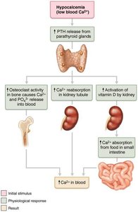
Antagonist to parathyroid hormone (PTH); inhibits osteoclast activity and stimulates Ca2+ uptake into bone.
Parathyroid Gland and Hormone
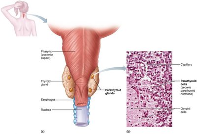
The parathyroid glands are small glands located on the posterior aspect of the thyroid. They secrete parathyroid hormone (PTH), which is the most important hormone in Ca2+ homeostasis.
Clinical Imbalances of Parathyroid Hormone
Hyperparathyroidism: Calcium leaches from bones, causing them to soften and deform; kidney stones may form.
Hypoparathyroidism: Can cause hypocalcemia, tetany, respiratory paralysis, and death.
Adrenal Glands
Structure and Function
The adrenal glands are paired, pyramid-shaped organs atop the kidneys. They consist of the adrenal cortex (three layers producing corticosteroids) and the adrenal medulla (producing catecholamines).
Adrenal Cortex Hormones
Mineralocorticoids (e.g., aldosterone): Regulate electrolyte concentrations.
Glucocorticoids (e.g., cortisol): Influence metabolism and help resist stressors.
Gonadocorticoids: Weak androgens contributing to puberty and sex drive.
Adrenal Medulla Hormones
Epinephrine and norepinephrine: Medullary chromaffin cells synthesize these catecholamines, which are involved in the fight-or-flight response.
Clinical Imbalances of Adrenal Hormones
Cushing's syndrome: Hypersecretion of glucocorticoids.
Addison's disease: Hyposecretion of glucocorticoids and mineralocorticoids.
Pheochromocytoma: Tumor causing hypersecretion of catecholamines.
Pineal Gland
Structure and Function
The pineal gland is a small gland hanging from the roof of the third ventricle. Pinealocytes secrete melatonin, which regulates day/night cycles and physiological rhythms.
Pancreas
Structure and Function
The pancreas is a triangular gland located behind the stomach. It has both exocrine (acinar cells) and endocrine (islets of Langerhans) functions.
Alpha cells: Produce glucagon (raises blood glucose).
Beta cells: Produce insulin (lowers blood glucose).
Clinical Imbalances of Pancreatic Hormones
Diabetes mellitus: Due to hyposecretion (Type 1) or hypoactivity (Type 2) of insulin.
Hyperinsulinism: Excessive insulin secretion causes hypoglycemia.
Gonads and Placenta
Structure and Function
Ovaries: Produce estrogens and progesterone; regulate reproductive organ maturation and secondary sexual characteristics.
Testes: Produce testosterone; initiates maturation of male reproductive organs and sex drive.
Placenta: Secretes estrogens, progesterone, and human chorionic gonadotropin (hCG).
Other Hormone-Producing Organs
Adipose tissue: Leptin, resistin, adiponectin.
Gastrointestinal tract: Gastrin, ghrelin, secretin, cholecystokinin, incretins.
Heart: Atrial natriuretic peptide (ANP).
Kidneys: Erythropoietin, renin.
Skeleton: Osteocalcin.
Skin: Cholecalciferol, calcitriol.
Thymus: Thymulin, thymopoietins, thymosins.
