 뒤로
The Kidneys: Structure, Function, and Physiology
스터디 가이드 - 스마트 노트
 자료에 맞춘 맞춤형 노트, 핵심 정의, 예시, 맥락을 확장해 제공합니다.
The Kidneys: Structure, Function, and Physiology
Overview
This chapter provides an in-depth exploration of the kidneys, their anatomical structure, physiological functions, and their critical role in maintaining homeostasis within the human body. The kidneys are essential organs in the urinary system, responsible for filtering blood, regulating fluid and electrolyte balance, and excreting waste products.
Functions of the Kidneys
Main Functions
Regulation of Extracellular Fluid Volume and Blood Pressure: The kidneys adjust the volume of blood plasma, influencing blood pressure.
Regulation of Osmolarity: They maintain the osmotic balance of body fluids.
Maintenance of Ion Balance: The kidneys regulate concentrations of key ions such as Na+, K+, and Ca2+.
Homeostatic Regulation of pH: By excreting or retaining H+ and HCO3-, the kidneys help maintain acid-base balance.
Excretion of Wastes: Metabolic wastes (e.g., urea, creatinine) and foreign substances are eliminated in urine.
Production of Hormones: The kidneys produce hormones such as erythropoietin (stimulates red blood cell production) and renin (regulates blood pressure).
Anatomy of the Urinary System
Components
Kidneys: Paired organs that filter blood and produce urine.
Ureters: Tubes that transport urine from the kidneys to the urinary bladder.
Urinary Bladder: Stores urine until it is excreted.
Urethra: Conducts urine from the bladder to the outside of the body. In males, it passes through the penis; in females, it opens anterior to the vagina.
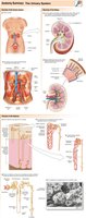
Kidney Structure
Location: Retroperitoneal, behind the peritoneum.
Renal Arteries and Veins: Supply and drain blood to and from the kidneys.
Nephron: The functional unit of the kidney, responsible for urine formation.
Cortex and Medulla: The outer cortex contains ~80% of nephrons (cortical nephrons); the inner medulla contains ~20% (juxtamedullary nephrons).
Vascular Elements: Blood flows through a portal system: afferent arteriole → glomerulus → efferent arteriole → peritubular capillaries/vasa recta.
Nephron Structure
Bowman's Capsule and Glomerulus: Site of plasma filtration, together called the renal corpuscle.
Proximal Tubule: Major site of reabsorption.
Loop of Henle: Has descending and ascending limbs; important for concentrating urine.
Distal Tubule and Collecting Ducts: Final adjustments to urine composition.
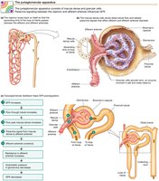
Juxtaglomerular Apparatus: Region where the ascending limb passes between afferent and efferent arterioles, involved in regulating blood pressure and filtration.
Overview of Kidney Function
Basic Processes
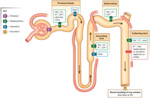
Filtration: Movement of fluid from blood into the nephron at the renal corpuscle. The filtered fluid is called filtrate.
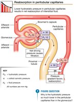
Reabsorption: Movement of substances from the filtrate back into the blood, primarily in the proximal tubule and peritubular capillaries.
Secretion: Active transport of substances from blood into the nephron tubule.
Excretion: Removal of substances from the body in urine.
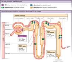
Modification of Filtrate
At the renal corpuscle, filtrate is nearly identical to plasma (180 L/day, 300 mOsm).
About 70% of filtrate is reabsorbed in the proximal tubule (solutes and water).
In the loop of Henle, more solute than water is reabsorbed, diluting the filtrate.
Distal tubule and collecting duct perform further reabsorption and secretion, leaving 1.5 L/day of urine (100–1200 mOsm, depending on hydration).
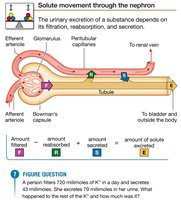
Filtration
Filtration Fraction and Barriers
Filtration Fraction: The percentage of renal plasma flow that becomes filtrate (typically ~20%).
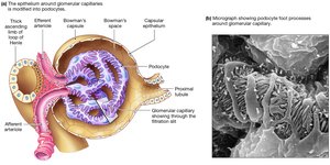
Filtration Barriers: The renal corpuscle has three filtration barriers:
Glomerular capillary endothelium (fenestrated capillaries and glycocalyx)
Basement membrane
Epithelium of Bowman's capsule (podocytes with filtration slits)
Mesangial Cells: Support glomerular capillaries and regulate filtration.
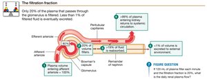
Forces Driving Filtration
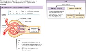
Capillary Blood Pressure (Hydrostatic Pressure): Favors filtration into Bowman's capsule.
Capillary Colloid Osmotic Pressure: Due to plasma proteins, opposes filtration.
Capsule Fluid Pressure: Hydrostatic pressure inside Bowman's capsule, opposes filtration.
Net Filtration Pressure: The sum of these forces determines the direction and rate of filtration.
Glomerular Filtration Rate (GFR)
Definition: The volume of fluid filtered per unit time (ml/min).
Determinants: Net filtration pressure, renal blood flow, blood pressure, and the filtration coefficient (surface area and permeability).
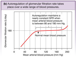
Regulation: GFR is kept relatively constant by autoregulation mechanisms, primarily by adjusting resistance in afferent and efferent arterioles.
Autoregulation of GFR
Myogenic Response: Vascular smooth muscle responds to pressure changes to maintain constant GFR.
Tubuloglomerular Feedback: Paracrine signaling at the juxtaglomerular apparatus adjusts arteriole resistance based on NaCl detection by macula densa cells.
Hormonal and Neural Control: Hormones and autonomic neurons can alter arteriole resistance and filtration coefficient.
Reabsorption
Mechanisms of Reabsorption
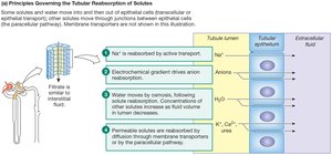
Transepithelial (Transcellular) Transport: Substances cross both apical and basolateral membranes of tubule epithelial cells.
Paracellular Pathway: Substances pass through cell–cell junctions between adjacent cells.
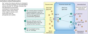
Active Transport: Sodium is actively transported, creating electrical and osmotic gradients that drive reabsorption of anions and water.
Secondary Active Transport: Glucose, amino acids, and other organics are reabsorbed via symport with sodium.
Passive Reabsorption: Urea and other solutes move by diffusion following concentration gradients.
Endocytosis: Plasma proteins are reabsorbed by receptor-mediated endocytosis.
Renal Transport Saturation
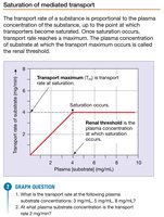
Saturation: Occurs when all transporters for a substance are occupied; transport rate reaches a maximum (transport maximum, Tm).
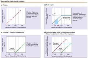
Renal Threshold: The plasma concentration at which a substance first appears in urine.
Example: Glucose is normally reabsorbed completely, but if plasma glucose exceeds the renal threshold, glucose appears in urine (glucosuria).
Secretion
Mechanisms and Importance
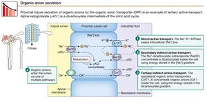
Definition: Active movement of molecules from extracellular fluid into the nephron lumen.
Homeostatic Role: Secretion helps regulate K+, H+, and organic ions, and increases excretion of substances.
Organic Anion Transporters (OATs): Transport a wide variety of substrates; competition can affect drug excretion (e.g., penicillin).
Excretion and Renal Clearance
Renal Handling and Clearance
Excretion: The removal of substances from the body in urine.
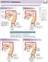
Clearance: The rate at which a solute disappears from the body by excretion or metabolism, expressed as volume of plasma cleared per minute.
Measurement: Inulin and creatinine are used to measure GFR. Inulin is freely filtered but neither reabsorbed nor secreted; creatinine is produced at a constant rate but is slightly secreted.
Useful Equations in Renal Physiology
Filtration Rate:
Excretion Rate:
Clearance:
Micturition
Urine Storage and Elimination
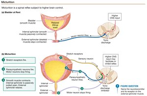
Urine Storage: Urine is stored in the bladder after leaving the collecting ducts; its composition does not change after this point.
Micturition Reflex: The process of urination is a spinal reflex controlled by two sphincters (internal and external). Stretch receptors in the bladder wall trigger the reflex, which can be overridden by higher brain centers.
Summary Table: Key Processes in the Nephron
Process | Location | Main Function |
|---|---|---|
Filtration | Renal corpuscle (glomerulus & Bowman's capsule) | Movement of plasma from blood to nephron |
Reabsorption | Proximal tubule, loop of Henle, distal tubule, collecting duct | Return of useful substances to blood |
Secretion | Proximal and distal tubules, collecting duct | Active transport of substances from blood to nephron |
Excretion | Collecting duct to ureter | Removal of waste in urine |
