 뒤로
The Muscle System: Structure, Function, and Major Muscles
스터디 가이드 - 스마트 노트
 자료에 맞춘 맞춤형 노트, 핵심 정의, 예시, 맥락을 확장해 제공합니다.
The Muscle System
Muscle Structure and Organization
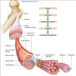
The muscular system is composed of specialized tissues that enable movement, maintain posture, and support bodily functions. Skeletal muscle is organized in a hierarchical structure, from the whole muscle down to the molecular level.
Muscle: The entire organ, attached to bones via tendons.
Fascicles: Bundles of muscle fibers within the muscle.
Muscle fibers (cells): Individual muscle cells, also called myocytes.
Myofibrils: Rod-like units within muscle fibers, composed of repeating sarcomeres.
Thick and thin filaments: Actin (thin) and myosin (thick) filaments responsible for contraction.
Connective tissue layers: Epimysium (surrounds muscle), perimysium (surrounds fascicles), endomysium (surrounds individual fibers).
Muscle Action and Body Movement
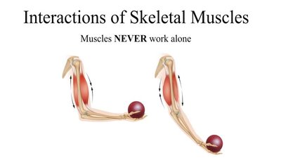
Muscle contraction is the basis for all voluntary movement. Muscles can only pull, not push, and work in groups to produce coordinated actions.
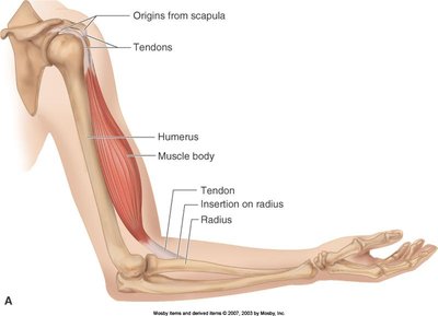
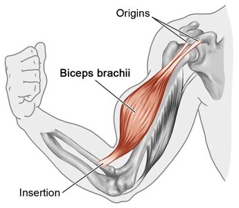
Origin: The stationary attachment point of a muscle during contraction.
Insertion: The movable attachment point; insertion moves toward origin during contraction.
Prime mover (agonist): Main muscle responsible for a movement.
Antagonist: Muscle that opposes or reverses the action of the prime mover.
Synergist: Assists the prime mover, adds force, or reduces unwanted movement.
Fixator: Stabilizes the origin of the prime mover.
Muscle Contraction: Factors Affecting Force and Movement
The force, velocity, and duration of muscle contraction depend on several factors:
Number of muscle fibers stimulated (recruitment): More motor units recruited increases force.
Relative size of fibers: Larger muscles generate more tension.
Frequency of stimulation: Higher frequency increases force.
Degree of muscle stretch: Optimal sarcomere length (80–120% of resting length) produces maximum force.
Load: Heavier loads slow contraction and reduce duration.
Muscle Fiber Types
Muscle fibers are classified by speed of contraction and metabolic properties:
Slow fibers: Endurance, fatigue-resistant.
Fast fibers: Powerful, fatigue quickly.
Effects of Exercise on Muscles
Aerobic (endurance) exercise: Increases capillaries, mitochondria, myoglobin; improves endurance and resistance to fatigue.
Resistance (anaerobic) exercise: Causes hypertrophy (fiber size increase), increases strength and muscle mass.
Disuse atrophy: Muscle degeneration due to inactivity or loss of neural stimulation.
Smooth Muscle Tissue
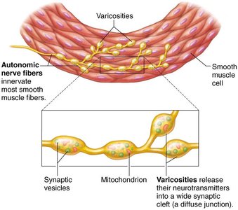
Smooth muscle is found in the walls of most hollow organs (except the heart). It is non-striated, involuntary, and regulated by the autonomic nervous system.
Structure: Two layers—longitudinal (shortens organ) and circular (constricts lumen).
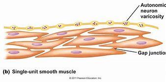
Varicosities: Bulbous nerve fiber swellings that release neurotransmitters into diffuse junctions.
Gap junctions: Electrically couple cells for synchronized contraction.
Contraction: Slow, sustained, and energy-efficient; can be self-excitatory.
Regulation: Controlled by nerves, hormones, or local chemicals; neurotransmitter effects vary by organ.
Fascicle Arrangement and Muscle Shape
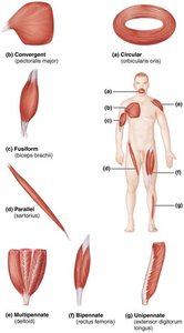
The arrangement of fascicles determines muscle shape and function:
Circular: Concentric rings (e.g., orbicularis oris).
Convergent: Broad origin, fascicles converge to a single tendon (e.g., pectoralis major).
Parallel: Fascicles run parallel to long axis (e.g., sartorius).
Pennate: Short fascicles attach obliquely to central tendon (e.g., rectus femoris).
Levers in the Muscular System
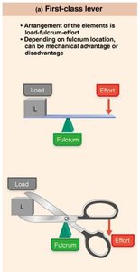
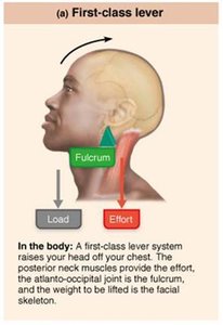
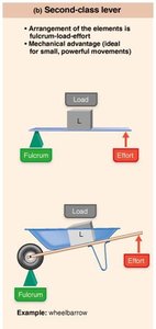
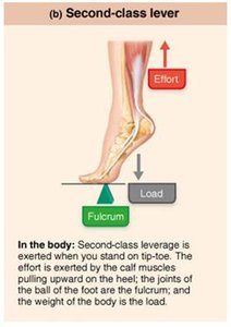
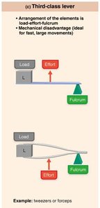
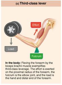
Most skeletal muscles use leverage to move bones. The lever system consists of:
Lever: Bone
Fulcrum: Joint
Effort: Force applied by muscle
Load: Resistance moved
Types of levers:
First-class lever: Fulcrum between load and effort (e.g., neck muscles raising head).
Second-class lever: Load between fulcrum and effort (e.g., standing on tiptoe).
Third-class lever: Effort between fulcrum and load (e.g., flexing forearm by biceps).
Major Muscles: Points of Attachment and Function
Muscles of the Head and Face
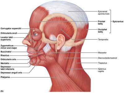
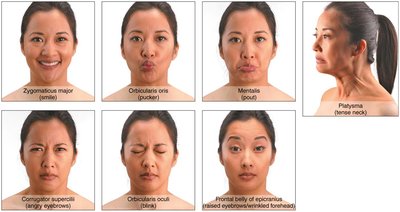
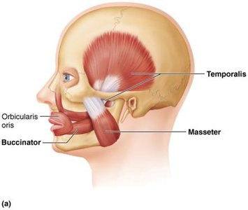
Muscles of facial expression insert into skin, enabling nonverbal communication. Muscles of mastication move the jaw for chewing.
Facial expression muscles: e.g., orbicularis oris, zygomaticus, platysma.
Mastication muscles: Temporalis, masseter (jaw closure); pterygoids (grinding); buccinator (chewing).
Tongue muscles: Genioglossus, hyoglossus, styloglossus (anchor and move tongue).
Neck and Back Muscles
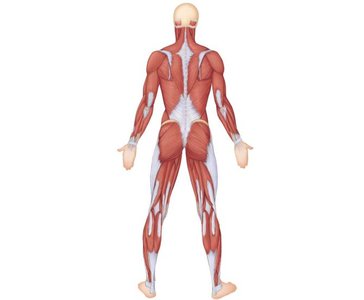
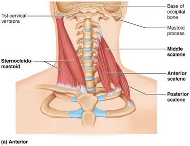
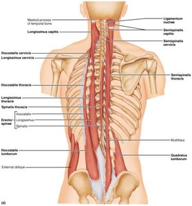
The sternocleidomastoid divides the neck into triangles and moves the head. Intrinsic back muscles (erector spinae, quadratus lumborum) extend the trunk and maintain posture.
Sternocleidomastoid: Flexes and rotates head.
Erector spinae group: Iliocostalis, longissimus, spinalis (extend vertebral column).
Quadratus lumborum: Lateral flexion of trunk.
Muscles of Breathing
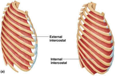
Breathing involves inspiratory and expiratory muscles.
Inspiratory muscles: Diaphragm, external intercostals (enlarge rib cage).
Expiratory muscles: Internal intercostals, abdominal muscles (decrease rib cage size).
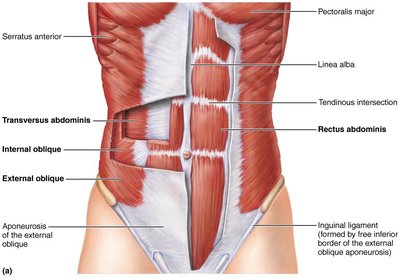
Abdominal Muscles
Four paired muscles form the lateral and anterior abdominal wall, enabling trunk movement and supporting visceral functions.
Rectus abdominis
External obliques
Internal obliques
Transversus abdominis
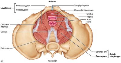
Pelvic Diaphragm Muscles
The pelvic diaphragm supports pelvic organs and resists intra-abdominal pressure.
Levator ani
Coccygeus
Shoulder and Arm Muscles
Extrinsic shoulder muscles fix and move the scapula, increasing arm movement range. The arm's prime movers include pectoralis major, latissimus dorsi, and deltoid. Rotator cuff muscles stabilize the shoulder.
Pectoralis major: Flexes, adducts arm.
Latissimus dorsi: Extends, adducts arm.
Deltoid: Abducts arm.
Rotator cuff: Supraspinatus, infraspinatus, teres minor, subscapularis.
Arm and Forearm Muscles
Arm muscles are divided into anterior (flexors) and posterior (extensors) compartments.
Biceps brachii: Flexes and supinates forearm.
Brachialis: Flexes forearm.
Brachioradialis: Flexes forearm.
Triceps brachii: Extends forearm.
Thigh Muscles
Thigh muscles are grouped by function: flexors, extensors, adductors, abductors, and rotators.
Thigh flexors: Iliopsoas, tensor fasciae latae, rectus femoris.
Thigh extensors: Hamstring group (biceps femoris, semitendinosus, semimembranosus).
Adductors: Adductor magnus, adductor longus, adductor brevis, pectineus, gracilis.
Abductors and rotators: Gluteus maximus, medius, minimus, piriformis, obturator externus/internus, gemellus.
Leg Muscles
Leg muscles control movements of the ankle, foot, and toes.
Anterior compartment: Tibialis anterior, extensor digitorum longus, extensor hallucis longus (dorsiflexion, toe extension).
Posterior compartment: Soleus, tibialis posterior, gastrocnemius (plantar flexion).
Summary Table: Major Muscle Groups and Functions
Muscle Group | Location | Function |
|---|---|---|
Facial Expression | Face | Nonverbal communication |
Mastication | Jaw | Chewing |
Neck | Neck | Head movement |
Back | Trunk | Posture, trunk extension |
Abdominal | Abdomen | Trunk flexion, visceral support |
Pelvic Diaphragm | Pelvis | Support pelvic organs |
Shoulder | Shoulder | Arm movement |
Arm | Upper limb | Forearm flexion/extension |
Thigh | Upper leg | Hip/knee movement |
Leg | Lower leg | Ankle/foot movement |
Key Equations
Force of muscle contraction: Where F is total force, n is number of cross bridges, f is force per cross bridge.
Mechanical advantage:
Additional info:
Muscle names often reflect location, shape, size, direction, number of origins, attachment points, or action.
Muscle health depends on regular activity; inactivity leads to atrophy.
