 뒤로
The Nervous System: Structure, Function, and Cellular Organization
스터디 가이드 - 스마트 노트
 자료에 맞춘 맞춤형 노트, 핵심 정의, 예시, 맥락을 확장해 제공합니다.
The Nervous System: Overview
Anatomical Divisions of the Nervous System
The nervous system is the primary controlling and communicating system of the body, responsible for integrating and responding to internal and external stimuli. It is divided into two main anatomical divisions:
Central Nervous System (CNS): Includes the brain and spinal cord. It processes information and directs responses.
Peripheral Nervous System (PNS): Consists of nerves that connect the CNS to the rest of the body, including cranial nerves, spinal nerves, and ganglia outside the CNS.
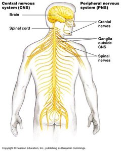
The CNS and PNS work together as a coordinated unit to maintain homeostasis and enable complex behaviors.
Functions of the Nervous System
The nervous system performs three interconnected functions:
Sensory Input: Specialized receptors receive stimuli from the environment.
Integration: The CNS processes and interprets sensory information.
Motor Output: The CNS sends signals to effector organs (muscles and glands) to produce a response.
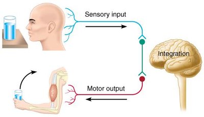
These functions allow the body to react to changes and maintain internal balance.
Organization of the Peripheral Nervous System (PNS)
Functional Subdivisions of the PNS
The PNS is organized into two main functional subdivisions:
Sensory (Afferent) Division: Transmits impulses from sensory receptors to the CNS. Includes somatic afferents (skin, muscles, joints), special sensory afferents, and visceral afferents (internal organs).
Motor (Efferent) Division: Transmits impulses from the CNS to effector organs. Includes motor neurons that activate muscles and glands.
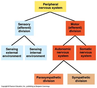
Motor Division: Somatic and Autonomic Nervous Systems
The motor portion of the PNS is further divided into:
Somatic Nervous System: Controls voluntary movements by activating skeletal muscles.
Autonomic Nervous System: Controls involuntary responses, regulating heart, smooth muscle, and glands. It has two divisions:
Sympathetic Division: Prepares the body for "fight or flight" emergencies.
Parasympathetic Division: Maintains "business as usual" functions such as digestion and rest.
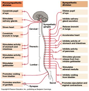
The two divisions often innervate the same organs but produce opposite effects.
Histology of Nervous Tissue
Cell Types in Nervous Tissue
Nervous tissue consists of two main functional cell types:
Neurons: Excitable cells that transmit electrical signals. They are specialized for communication and have extreme longevity, are amitotic, and require a constant supply of oxygen and glucose.
Neuroglia (Glial Cells): Support cells that provide structural and metabolic support to neurons.
Neurons are the functional units of the nervous system, transmitting signals via electrical charges across their membranes.
Structure of a Neuron
Neurons have three major parts:
Cell Body (Soma): Contains the nucleus and organelles; site of synthesis and metabolism.
Dendrites: Short, highly branched processes that receive and transmit signals to the cell body.
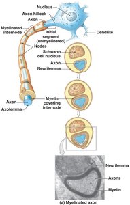
Axon: Long, thin projection that conveys information away from the soma to other neurons or effector cells. Axons may be covered by a myelin sheath formed by neuroglia.
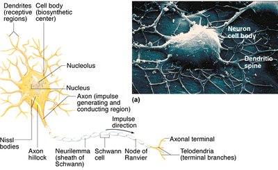
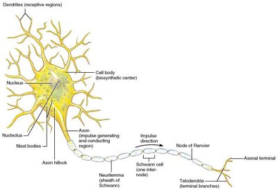
Clusters of neuron cell bodies are called nuclei in the CNS and ganglia in the PNS.
Classification of Neurons
Structural Classification
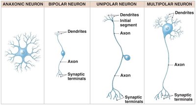
Neurons are classified based on the number of processes extending from the cell body:
Anaxonic Neurons: Found in the brain and sensory organs.
Bipolar Neurons: Found in special sensory organs (sight, smell, hearing).
Unipolar Neurons: Found in the sensory part of the PNS.
Multipolar Neurons: Common in the CNS; include all skeletal muscle motor neurons.
Functional Classification
Neurons are also classified by the direction of nerve impulses:
Sensory (Afferent) Neurons: Transmit impulses from sensory receptors to the CNS.
Motor (Efferent) Neurons: Transmit impulses from the CNS to muscles or glands.
Interneurons (Association Neurons): Convey impulses between parts of the CNS; involved in higher functions such as memory, planning, and learning.
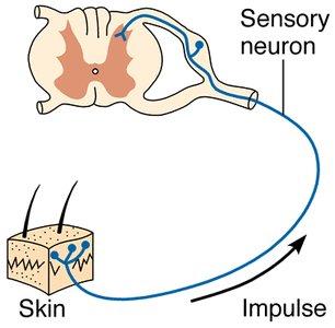
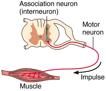
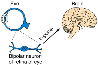
Synapses and Signal Transmission
Types of Synapses
Neurons communicate at specialized junctions called synapses:
Neuro-neural Junction: Between two neurons.
Neuro-muscular Junction: Between a neuron and a muscle.
Neuro-glandular Junction: Between a neuron and a gland.
The synapse includes:
Presynaptic Cell: Sends the message.
Postsynaptic Cell: Receives the message.
Synaptic Cleft: Small gap separating the presynaptic and postsynaptic membranes.
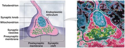
Neuroglia: Support Cells of the Nervous System
Types of Neuroglia in the CNS
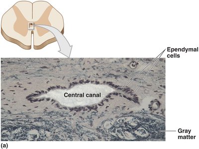
Ependymal Cells: Form the ependyma, line the central canal of the spinal cord and ventricles of the brain, secrete and circulate cerebrospinal fluid (CSF), and contain stem cells for repair.
Microglia: Migrate through neural tissue and act as phagocytes, cleaning up debris and pathogens.
Astrocytes: Maintain the blood–brain barrier, provide structural support, repair damaged tissue, guide neuron development, and control the interstitial environment.
Oligodendrocytes: Wrap around axons to form myelin sheaths in the CNS.
Types of Neuroglia in the PNS
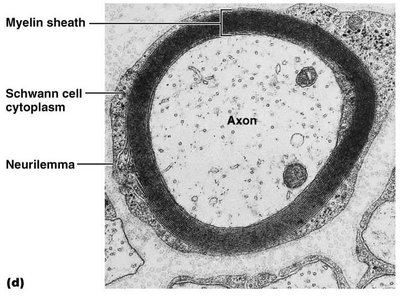
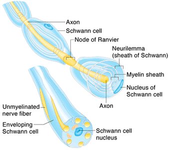
Schwann Cells (Neurolemmocytes): Form the myelin sheath around axons in the PNS. The gaps between Schwann cells are called nodes of Ranvier.
Myelination and Nerve Fiber Types
Myelinated vs. Unmyelinated Axons
Myelination occurs during fetal development and the first year of life. Myelinated fibers transmit impulses faster than unmyelinated fibers. Schwann cells provide a path for regrowth of neuron processes after injury in the PNS.
Gray Matter: Unmyelinated cell bodies and processes in the CNS.
White Matter: Myelinated processes in various fiber tracts in the CNS.
Summary Table: Structural and Functional Classification of Neurons
Classification | Structural Types | Functional Types |
|---|---|---|
Location | Anaxonic, Bipolar, Unipolar, Multipolar | Sensory (afferent), Motor (efferent), Interneuron |
Example | Bipolar (retina), Multipolar (motor neuron) | Sensory neuron (skin), Motor neuron (muscle), Interneuron (CNS) |
Key Function | Number and arrangement of processes | Direction of impulse transmission |
Key Equations and Concepts
Action Potential: The electrical signal transmitted along the axon. Initiated at the axon hillock (trigger zone).
Resting Membrane Potential:
Impulse Transmission Speed: Myelinated fibers transmit impulses faster due to saltatory conduction at nodes of Ranvier.
Example: The sympathetic division increases heart rate during emergencies, while the parasympathetic division slows it during rest.
Additional info: Academic context was added to clarify neuron structure, synaptic function, and neuroglia roles.
