 뒤로
The Respiratory System: Structure and Function
스터디 가이드 - 스마트 노트
 자료에 맞춘 맞춤형 노트, 핵심 정의, 예시, 맥락을 확장해 제공합니다.
The Respiratory System
Overview and Major Functions
The respiratory system is essential for gas exchange, supplying oxygen (O2) to body tissues and removing carbon dioxide (CO2) produced by cellular metabolism. It works closely with the cardiovascular system to ensure efficient transport and exchange of respiratory gases. Additional functions include olfaction (sense of smell) and speech production.
Gas Exchange: Supplies O2 to cells and removes CO2.
Olfaction: Detects odors via olfactory receptors in the nasal cavity.
Speech: Air movement through the larynx enables vocalization.
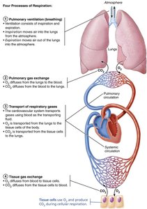
Four Processes of Respiration
Pulmonary Ventilation (Breathing): Movement of air into and out of the lungs.
Pulmonary Gas Exchange: Exchange of O2 and CO2 between alveoli and blood.
Transport of Respiratory Gases: Movement of O2 and CO2 in the blood between lungs and tissues.
Tissue Gas Exchange: Exchange of O2 and CO2 between blood and body tissues.
Functional Anatomy of the Respiratory System
Major Organs and Divisions
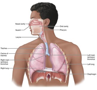
The respiratory system is divided into the upper respiratory system (nose, paranasal sinuses, pharynx) and the lower respiratory system (larynx, trachea, bronchi, lungs, and alveoli). Respiratory muscles, such as the diaphragm and intercostal muscles, are essential for ventilation.
Upper Respiratory System
Nose (External Nose and Nasal Cavity): Provides airway, moistens/warms/filters air, resonance for speech, houses olfactory receptors.
Paranasal Sinuses: Lighten skull, warm/moisten/filter air, secrete mucus.
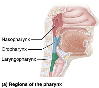
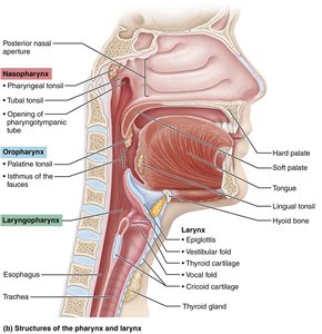
Pharynx: Passageway for air and food; houses tonsils for immune defense.
Structure | Description | Function |
|---|---|---|
Nose | Supported by bone and cartilage; lined with mucosa; contains olfactory epithelium | Filters, warms, moistens air; resonance for speech; olfaction |
Paranasal Sinuses | Mucosa-lined, air-filled cavities in cranial bones | Lighten skull; warm/moisten/filter air |
Pharynx | Connects nasal cavity to larynx and oral cavity to esophagus; three regions | Passageway for air/food; immune defense |
The Nose and Paranasal Sinuses
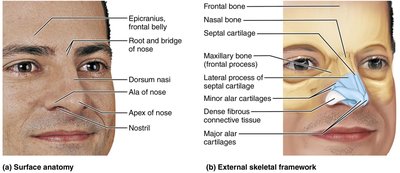
External Nose: Formed by nasal/frontal bones, maxillary bones, and hyaline cartilage. Surface features include root, bridge, dorsum nasi, apex, and nostrils (nares).
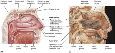
Nasal Cavity: Divided by the nasal septum; lined with mucosa (olfactory and respiratory). Contains nasal conchae to increase surface area and turbulence for air filtration.
Paranasal Sinuses: Located in frontal, sphenoid, ethmoid, and maxillary bones; drain into nasal cavity.
The Pharynx
Nasopharynx: Airway posterior to nasal cavity; contains pharyngeal tonsils and auditory tubes.
Oropharynx: Posterior to oral cavity; passageway for food and air; contains palatine and lingual tonsils.
Laryngopharynx: Posterior to larynx; passageway for food and air; continuous with esophagus.
Lower Respiratory System
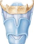
Larynx (Voice Box): Provides open airway, routes air/food, voice production; composed of nine cartilages (thyroid, cricoid, epiglottis, arytenoid, cuneiform, corniculate).
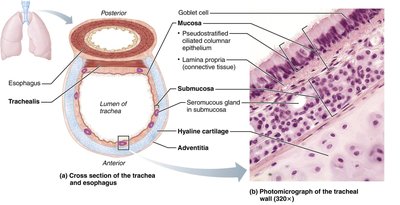
Trachea (Windpipe): Flexible tube with C-shaped cartilage rings; conducts, cleans, warms, and moistens air.
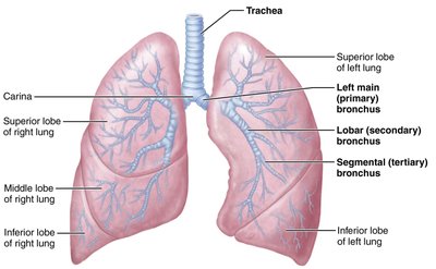
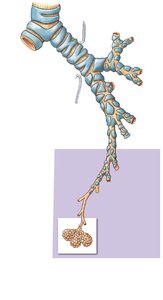
Bronchial Tree: Branching system of bronchi and bronchioles; conducts air to alveoli.
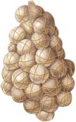
Alveoli: Microscopic air sacs; main sites of gas exchange; produce surfactant to reduce surface tension.
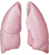
Lungs: Paired organs composed of alveoli and elastic connective tissue; divided into lobes and bronchopulmonary segments.
Pleurae: Serous membranes (parietal and visceral) that produce lubricating fluid and compartmentalize lungs.
Structure | Description | Function |
|---|---|---|
Larynx | Cartilaginous structure connecting pharynx to trachea | Air passageway; prevents food entry; voice production |
Trachea | Flexible tube with C-shaped cartilage rings | Air passageway; cleans, warms, moistens air |
Bronchial Tree | Branching airways from trachea to alveoli | Conducts, cleans, warms, moistens air |
Alveoli | Microscopic air sacs | Main sites of gas exchange; surfactant production |
Lungs | Paired, elastic organs | House respiratory passages; gas exchange |
Pleurae | Serous membranes | Lubrication; compartmentalization |
Histology and Microscopic Structure
Tracheal Wall Structure
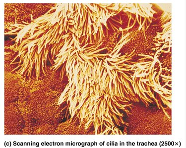
Mucosa: Pseudostratified ciliated columnar epithelium with goblet cells (mucus production).
Submucosa: Connective tissue with seromucous glands.
Hyaline Cartilage: C-shaped rings prevent collapse.
Adventitia: Outermost connective tissue layer.
Conducting and Respiratory Zones
Conducting Zone
The conducting zone includes all respiratory passageways from the nose to the terminal bronchioles. Its primary function is to transport, filter, warm, and humidify air, but no gas exchange occurs here.
Respiratory Zone
The respiratory zone consists of respiratory bronchioles, alveolar ducts, and alveoli. This is the site of actual gas exchange between air and blood.
Summary Table: Upper vs. Lower Respiratory System
Region | Main Structures | Main Functions |
|---|---|---|
Upper Respiratory System | Nose, nasal cavity, paranasal sinuses, pharynx | Filter, warm, moisten air; olfaction; resonance for speech |
Lower Respiratory System | Larynx, trachea, bronchi, bronchioles, alveoli, lungs | Conduct air; gas exchange; voice production |
Key Terms and Concepts
Pulmonary Ventilation: Movement of air into and out of the lungs (breathing).
Gas Exchange: Diffusion of O2 and CO2 across respiratory membranes.
Surfactant: Substance produced by alveolar cells to reduce surface tension and prevent alveolar collapse.
Respiratory Membrane: Thin barrier (alveolar and capillary walls) where gas exchange occurs.
Pleura: Double-layered serous membrane surrounding each lung (parietal and visceral layers).
Clinical Connections
Rhinitis: Inflammation of nasal mucosa, leading to congestion and postnasal drip.
Laryngitis: Inflammation of vocal folds, causing hoarseness or loss of voice.
Tracheal Obstruction: Life-threatening; Heimlich maneuver can expel blockage.
Pleurisy: Inflammation of pleurae, causing pain and impaired breathing.
Summary
The respiratory system is a complex network of structures and tissues that work together to ensure efficient gas exchange, protect the body from pathogens, and facilitate communication. Understanding its anatomy and physiology is essential for diagnosing and treating respiratory disorders.
