 뒤로
The Skeletal System: Structure, Function, and Anatomy
스터디 가이드 - 스마트 노트
 자료에 맞춘 맞춤형 노트, 핵심 정의, 예시, 맥락을 확장해 제공합니다.
The Skeletal System
Overview of Mobility and Skeletal System Components
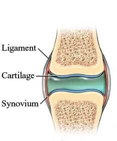
The skeletal system is essential for mobility, providing the framework for purposeful physical movement. Optimal mobility depends on the coordinated function of bones, joints, articular cartilage, tendons, ligaments, and skeletal muscle. Changes in mobility can significantly impact overall health, and healthcare professionals play a crucial role in promoting and maintaining optimal mobility.
Bones: Provide structure and support.
Joints: Allow movement between bones.
Cartilages: Cushion and support joints.
Ligaments: Connect bones and stabilize joints.
Functions of the Skeletal System
The skeletal system performs several critical functions necessary for life and health:
Support: Provides the structural framework for the body.
Protection: Shields vital organs (e.g., skull protects the brain, ribs protect the heart and lungs).
Movement: Acts as anchorage for muscles, enabling movement.
Storage: Stores minerals (calcium, phosphorus), growth factors, and triglycerides.
Hormone Production: Osteocalcin regulates bone formation and insulin secretion.
Blood Cell Formation (Hematopoiesis): Occurs in bone marrow.
Organization of the Skeletal System
Axial Skeleton
The axial skeleton forms the central axis of the body and consists of the skull, vertebral column, and thoracic cage.
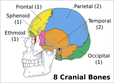
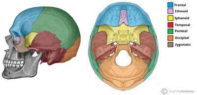
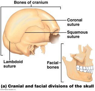
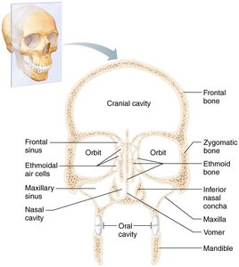
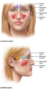
Skull: Composed of cranial and facial bones.
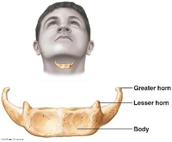
Hyoid Bone: Located in the neck, supports the tongue.
Vertebral Column: Protects the spinal cord and supports the head.
Thoracic Cage: Includes ribs and sternum, protects thoracic organs.
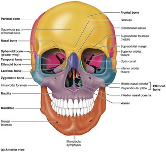
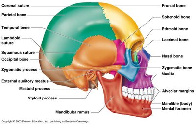
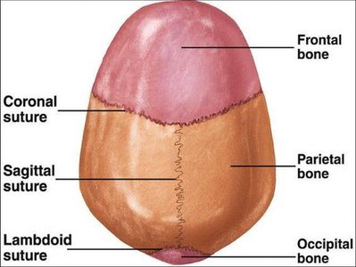
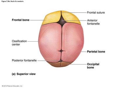
Thoracic Cage
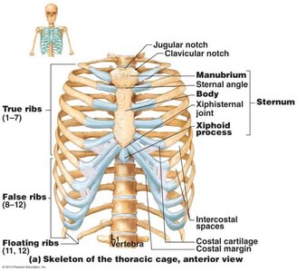
The thoracic cage consists of the ribs, sternum, and thoracic vertebrae. It protects the heart and lungs and supports the upper limbs.
Sternum: Divided into manubrium, body, and xiphoid process.
Ribs: Classified as true, false, and floating ribs.
Vertebral Column
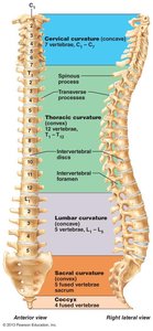
The vertebral column is composed of 33 vertebrae grouped into cervical, thoracic, lumbar, sacral, and coccygeal regions. It provides structural support and flexibility.
Cervical: 7 vertebrae
Thoracic: 12 vertebrae
Lumbar: 5 vertebrae
Sacral: 5 fused vertebrae
Coccyx: 4 fused vertebrae
Appendicular Skeleton
The appendicular skeleton includes the pectoral girdle, upper limbs, pelvic girdle, and lower limbs. It facilitates movement and interaction with the environment.
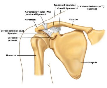
Pectoral Girdle: Clavicle and scapula
Upper Limbs: Humerus, radius, ulna, carpals, metacarpals, phalanges
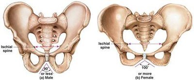
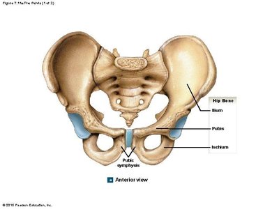
Pelvic Girdle: Ilium, ischium, pubis
Lower Limbs: Femur, tibia, fibula, tarsals, metatarsals, phalanges
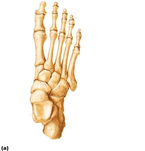
Classification of Bones by Shape
Types of Bones
Bones are classified by their shape, which relates to their function:
Long Bones: Longer than they are wide (e.g., humerus, femur)
Short Bones: Cube-shaped (e.g., carpals, tarsals)
Flat Bones: Thin, flattened, and usually curved (e.g., sternum, skull)
Irregular Bones: Complex shapes (e.g., vertebrae, hip bones)
Bone Structure and Anatomy
Gross Anatomy
Bones have two main textures:
Compact Bone: Dense outer layer, smooth and solid.
Spongy Bone (Trabecular Bone): Honeycomb structure with trabeculae.
Structure of Short, Irregular, and Flat Bones
These bones consist of thin plates of spongy bone (diploë) covered by compact bone, with periosteum and endosteum membranes. Bone marrow is present throughout spongy bone, and hyaline cartilage covers articular surfaces.
Structure of Typical Long Bone
Diaphysis: Tubular shaft, compact bone surrounds medullary cavity.
Epiphyses: Bone ends, external compact bone, internal spongy bone, articular cartilage covers surfaces.
Membranes: Periosteum (outer), endosteum (inner).
Epiphyseal Line: Remnant of childhood bone growth.
Membranes
Periosteum: White, double-layered, covers external surfaces except joints, contains osteogenic cells, nerve fibers, and blood vessels.
Endosteum: Covers internal bone surfaces, lines medullary cavity and canals, contains osteogenic cells.
Hematopoietic Tissue in Bones
Red Marrow: Found in trabecular cavities of spongy bone and diploë of flat bones; active in hematopoiesis.
Yellow Marrow: Can convert to red marrow if necessary.
Bone Markings
Bone markings are projections, surfaces, and depressions/openings that serve as sites for muscle, ligament, and tendon attachment, joint formation, and passage for blood vessels and nerves.
Projections: Tuberosity, crest, trochanter, line, tubercle, epicondyle, spine, process.
Surfaces: Head, facet, condyle.
Depressions/Openings: Groove, fissure, foramen, notch, meatus, sinus, fossa.
Chemical Composition of Bone
Organic Components
Osteogenic Cells: Stem cells that differentiate into osteoblasts.
Osteoblasts: Bone-forming cells, secrete osteoid (organic matrix).
Osteocytes: Mature bone cells, maintain bone matrix.
Bone-Lining Cells: Maintain matrix on bone surfaces.
Osteoclasts: Bone-resorbing cells, break down bone tissue.
Osteoid: Contains ground substance and collagen fibers, provides tensile strength and flexibility.
Inorganic Components
Hydroxyapatites (Mineral Salts): Mainly calcium phosphate crystals, responsible for bone hardness.
Microscopic Anatomy of Bone
Cells of Bone Tissue
Osteogenic Cells: Mitotically active, differentiate into osteoblasts or bone-lining cells.
Osteoblasts: Actively mitotic, secrete unmineralized bone matrix.
Osteocytes: Mature bone cells in lacunae, monitor and maintain bone matrix.
Bone-Lining Cells: Flat cells on bone surfaces, maintain matrix.
Osteoclasts: Derived from hematopoietic stem cells, giant multinucleate cells for bone resorption.
Compact Bone
Osteon (Haversian System): Structural unit, elongated cylinder parallel to bone axis.
Lamellae: Hollow tubes of bone matrix, collagen fibers in adjacent rings run in different directions.
Central (Haversian) Canal: Contains blood vessels and nerves.
Perforating (Volkmann's) Canal: Connects blood and nerve supply of periosteum to central canal.
Lacunae: Spaces containing osteocytes.
Canaliculi: Tiny canals connecting lacunae.
Spongy Bone
Trabeculae: Align along lines of stress, contain irregularly arranged lamellae, capillaries supply nutrients.
No Osteons: Structure is less organized than compact bone.
Bone Development and Growth
Ossification (Osteogenesis)
Ossification is the process of bone tissue formation, beginning in the second month of development. The embryonic skeleton consists of hyaline cartilage or fibrous membrane before becoming bone.
Endochondral Ossification: Bone forms by replacing hyaline cartilage; forms most of the skeleton.
Intramembranous Ossification: Bone develops from fibrous membrane; forms clavicles and cranial bones.
Postnatal Bone Growth
Interstitial Growth: Increases length of long bones; epiphyseal plate provides for growth.
Appositional Growth: Increases bone thickness; occurs throughout life.
Hormonal Regulation of Bone Growth
Growth Hormone: Stimulates epiphyseal plate activity in infancy and childhood.
Thyroid Hormone: Modulates activity of growth hormone.
Sex Hormones: Promote adolescent growth spurts and induce epiphyseal plate closure.
Bone Homeostasis and Remodeling
Bone Remodeling
Bone remodeling involves bone resorption and bone deposit, maintaining calcium homeostasis and responding to mechanical stress.
Bone Resorption: Osteoclasts break down bone, releasing calcium and growth factors into blood.
Bone Deposit: Osteoblasts lay down new bone matrix.
Remodeling: Allows for normal bone proportion and strength as body changes.
Calcium Homeostasis
Blood calcium levels are tightly regulated by hormonal control, primarily by parathyroid hormone (PTH).
Falling Blood Ca2+ Levels: Parathyroid glands release PTH, stimulating osteoclasts to degrade bone matrix and release Ca2+ into blood.
Rising Blood Ca2+ Levels: Thyroid gland releases calcitonin, inhibiting osteoclast activity.
Equation:
Bone Injury and Repair
Fractures and Bone Repair
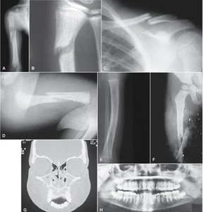
Fractures are breaks or cracks in bones, classified as closed/simple or open/compound. Bone repair occurs in four steps:
Hematoma Formation: Blood vessels break, forming a hematoma.
Fibrocartilage Callus Formation: New capillaries grow, fibroblasts secrete collagen, chondroblasts secrete cartilage.
Bony Callus Formation: Fibrocartilage callus replaced by spongy bone.
Bone Remodeling: Bony callus remodeled in response to stress, forming a strong permanent patch.
Cartilage: Structure, Types, and Locations
Basic Structure of Cartilage
Cartilage is a resilient connective tissue with no blood vessels or nerves. The perichondrium surrounds cartilage, providing nutrients and resisting expansion.
Chondrocytes: Cells in lacunae within the extracellular matrix.
Types of Cartilage
Hyaline Cartilage: Provides support, flexibility, and resilience; most abundant type.
Elastic Cartilage: Similar to hyaline but contains elastic fibers.
Fibrocartilage: Thick collagen fibers, great tensile strength.
Locations: External ear, intervertebral discs, pubic symphysis, meniscus, articular cartilage of joints, costal cartilage, nose, epiglottis, larynx, trachea.
