 뒤로
Urinary System: Structure, Function, and Pathology
스터디 가이드 - 스마트 노트
 자료에 맞춘 맞춤형 노트, 핵심 정의, 예시, 맥락을 확장해 제공합니다.
Urinary System Overview
General Structure and Function
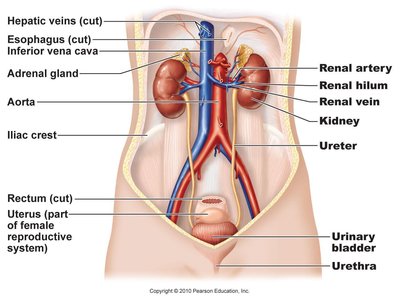
The urinary system is responsible for filtering blood, removing waste, and maintaining homeostasis of water, electrolytes, and acid-base balance. It consists of the kidneys, ureters, urinary bladder, and urethra.
Kidneys: Filter blood and produce urine.
Ureters: Transport urine from kidneys to bladder.
Urinary bladder: Stores urine until excretion.
Urethra: Conducts urine out of the body.

Kidney Anatomy and Location
External and Internal Structure
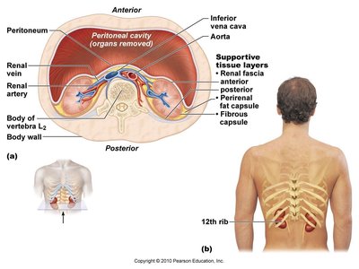
The kidneys are retroperitoneal organs located between T12 and L3 vertebrae, protected by an adipose capsule. Each kidney is about the size of a bar of soap.
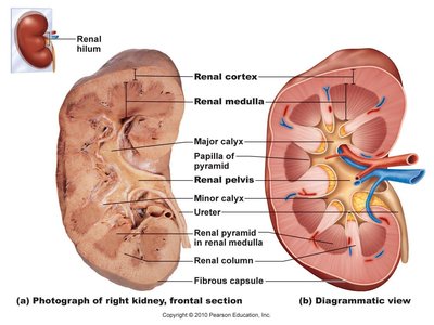
Renal Cortex: Outer region, contains nephrons.
Renal Medulla: Inner region, contains renal pyramids.
Renal Pelvis: Funnel-shaped structure collecting urine.
Renal Artery and Vein: Blood supply to and from the kidney.
Ureter: Exits at the renal pelvis.
Kidney Functions
Homeostatic Roles
The kidneys perform several essential functions:
Filtration: Removes waste from blood plasma.
Regulation: Controls osmolarity, blood volume, blood pressure, and acid-base balance.
Secretion: Produces hormones such as renin (regulates blood pressure) and erythropoietin (stimulates red blood cell production).
Detoxification: Metabolizes drugs and free radicals.
Nephron Structure and Function
Functional Unit of the Kidney
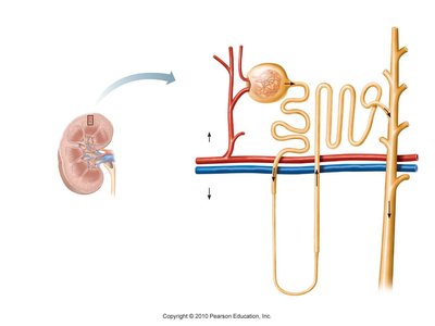
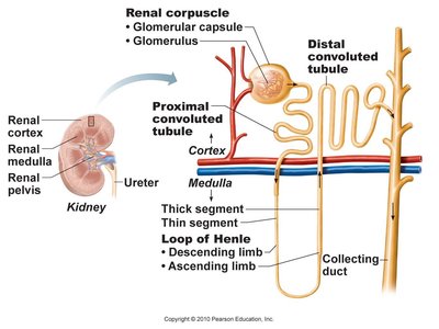
Nephrons are the microscopic units that form urine. Each kidney contains about 1.2 million nephrons.
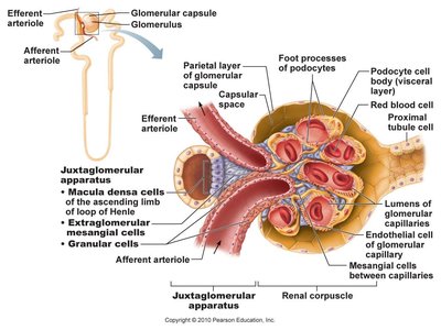
Renal Corpuscle: Includes the glomerulus and Bowman's capsule, where filtration occurs.
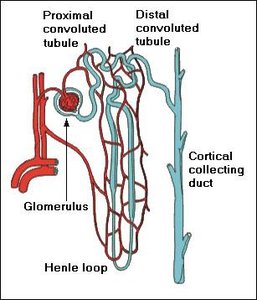
Renal Tubule: Includes the proximal convoluted tubule (PCT), nephron loop (Loop of Henle), distal convoluted tubule (DCT), and collecting duct (CD).
Cortical Nephrons: 85%, short loops, close to surface.
Juxtamedullary Nephrons: 15%, long loops, deep in medulla.
Urine Formation
Three Main Processes
Urine is formed through three processes:
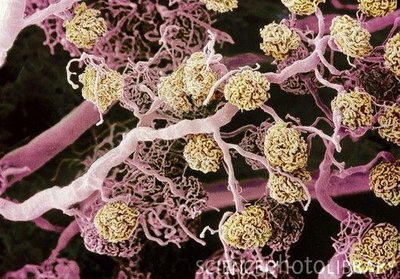
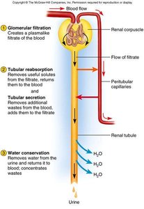
Glomerular Filtration: Blood pressure forces plasma through the filtration membrane in the glomerulus.
Tubular Reabsorption: Useful substances are reabsorbed from the filtrate back into the blood.
Tubular Secretion: Additional wastes are secreted from blood into the filtrate.
Filtration Membrane
The filtration membrane consists of:
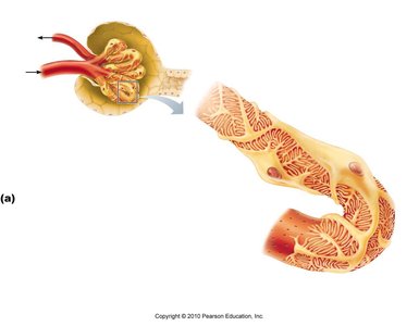
Fenestrated Endothelium: Allows passage of small molecules, excludes blood cells.
Basement Membrane: Excludes large molecules.
Filtration Slits: Podocyte arms allow only small particles to pass.
Filtration Pressure
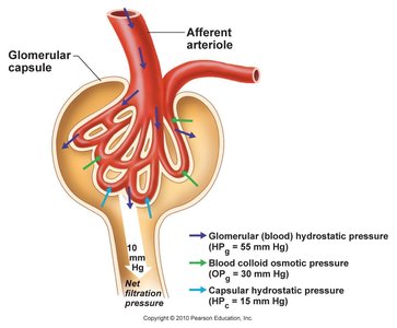
Filtration is driven by high blood pressure in the glomerulus, maintained by the wider afferent arteriole compared to the efferent arteriole.
Net Filtration Pressure:
Where HPg = glomerular hydrostatic pressure, OPg = blood colloid osmotic pressure, HPc = capsular hydrostatic pressure.
Glomerular Filtration Rate (GFR)
Definition and Clinical Importance
GFR: Volume of filtrate formed per minute by both kidneys.
Normal values: 125 ml/min (male), 105 ml/min (female).
99% of filtrate is reabsorbed; only 1-2 L urine excreted/day.
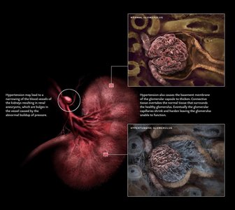
High GFR leads to dehydration and electrolyte loss; low GFR leads to anuria and waste retention.
Regulation of Glomerular Filtration
Mechanisms of Control
GFR is regulated by three mechanisms:
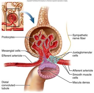
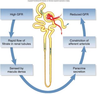
Autoregulation: Myogenic mechanism and tubuloglomerular feedback stabilize GFR.
Sympathetic Control: Constriction of afferent arterioles during stress reduces GFR.
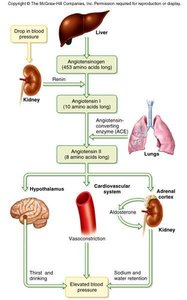
Hormonal Control: Renin-angiotensin-aldosterone system adjusts GFR and blood pressure.
Tubular Reabsorption and Secretion
Reabsorption in the Proximal Convoluted Tubule (PCT)
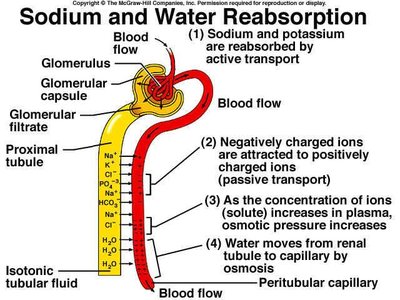
Most reabsorption occurs in the PCT, returning water, glucose, amino acids, and vitamins to the blood.
Peritubular Capillaries: Low blood pressure and high osmolarity favor reabsorption.
Solvent Drag: Water movement carries solutes with it.
Tubular Secretion
Tubular secretion removes wastes (urea, uric acid, ammonia, drugs) and regulates acid-base balance by secreting H+ and bicarbonate ions.
Nephron Loop (Loop of Henle)
Descending Limb: Reabsorbs water, not electrolytes.
Ascending Limb: Reabsorbs electrolytes (Na+, Cl-, K+), not water.
Creates a salinity gradient for urine concentration.
DCT and Collecting Duct
Fluid reabsorption regulated by hormones (aldosterone, ADH).
Maintains salt/water and acid/base balance.
Urine Concentration
Medullary collecting duct is highly permeable to water, allowing concentration of urine.
Osmolarity of interstitial fluid increases deep in the medulla.
Hormonal Effects
Aldosterone: Promotes Na+ and water reabsorption, decreases urine volume, increases blood pressure.
ADH: Increases water reabsorption, decreases urine volume.
Urinalysis and Properties of Urine
Physical and Chemical Characteristics
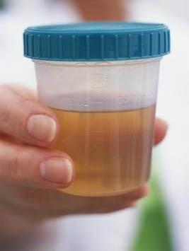
Appearance: Color ranges from odorless to deep amber; yellow due to urochrome.
Odor: Bacteria degrade urea to ammonia; certain foods affect smell.
Specific Gravity: Correlates to urine osmolarity.
pH: Range 4.5–8.2, usually 6.0.
Chemical Composition: 95% water, 5% solutes (urea, NaCl, KCl, creatinine, uric acid).
Nitrogenous Wastes
Urea: From amino acid metabolism.
Uric Acid: From nucleic acid catabolism.
Creatinine: From creatine phosphate catabolism.
Urine Volume and Disorders
Normal: 1–2 L/day.
Polyuria: >2 L/day.
Oliguria: <500 mL/day.
Anuria: 0–100 mL/day.
Diabetes Mellitus: Chronic polyuria, hyperglycemia, glycosuria.
Diabetes Insipidus: ADH hyposecretion.
Diuretics: Increase urine output, decrease blood volume.
Ureters, Bladder, and Urethra
Ureters
Transport urine from renal pelvis to bladder via peristalsis.
About 25 cm long.
Urinary Bladder
Highly distensible, capacity 500–800 mL.
Trigone: triangular area formed by ureter and urethra openings, susceptible to infection.
Rugae: folds in relaxed bladder.
Urethra
Female: 3–4 cm, more prone to infection.
Male: 18 cm.
Internal sphincter: involuntary control.
External sphincter: voluntary control.
Micturition (Voiding Urine)
Process and Control
Stretch receptors in bladder signal micturition center.
Results in bladder contraction and relaxation of internal sphincter.
Voluntary control via external sphincter and cerebrum.
Normal and Abnormal Constituents of Urine
Normal Constituents
Urea, creatinine, uric acid, urobilinogen.
Abnormal Constituents
Albumin (albuminuria), glucose (glucosuria), blood (hematuria), pus (pyuria), ketones (ketonuria), bilirubin (bilirubinuria), kidney stones, microbes (UTI).
Kidney Pathology
Common Disorders
Trauma, Infection, Strenuous Exercise, Prolonged Hypoxia: Can cause proteinuria or hematuria.
Polycystic Kidney Disease: Genetic disorder causing multiple cysts, chronic infections, kidney failure.
Kidney Stones: Renal calculi of calcium, phosphate, uric acid, protein; cause pain and obstruction.
Renal Insufficiency: Inability to maintain homeostasis due to nephron destruction.
Hemodialysis: Artificial filtration of blood for renal failure.
Renal Cell Carcinoma: Cancer of kidney tubule lining; symptoms include pain, abnormal urine color, swelling, weight loss.
Summary Table: Urine Formation Steps
Step | Location | Main Function |
|---|---|---|
Glomerular Filtration | Glomerulus | Filters plasma, forms filtrate |
Tubular Reabsorption | PCT, Loop of Henle, DCT, CD | Returns water and solutes to blood |
Tubular Secretion | PCT, DCT | Removes wastes, regulates pH |
Water Conservation | Collecting Duct | Concentrates urine |
Summary Table: Kidney Pathologies
Pathology | Cause | Effect |
|---|---|---|
Polycystic Kidney Disease | Genetic | Cysts, infection, failure |
Kidney Stones | Hypercalcemia, dehydration | Pain, obstruction |
Renal Insufficiency | Nephron destruction | Homeostasis failure |
Renal Cell Carcinoma | Unknown | Cancer, pain, abnormal urine |
Key Terms and Definitions
Nephron: Functional unit of the kidney.
Glomerulus: Capillary bed for filtration.
Bowman's Capsule: Surrounds glomerulus, collects filtrate.
Proximal Convoluted Tubule (PCT): Reabsorbs nutrients.
Loop of Henle: Creates salinity gradient.
Distal Convoluted Tubule (DCT): Regulates reabsorption/secretion.
Collecting Duct (CD): Concentrates urine.
Glomerular Filtration Rate (GFR): Rate of plasma filtration.
Renin-Angiotensin-Aldosterone System: Hormonal regulation of BP and GFR.
Additional info: Academic context was added to clarify nephron types, urine formation steps, and kidney pathologies for completeness.
