 뒤로
뒤로Comprehensive Study Notes on Carbohydrates for Biochemistry
스터디 가이드 - 스마트 노트
 자료에 맞춘 맞춤형 노트, 핵심 정의, 예시, 맥락을 확장해 제공합니다.
Carbohydrates: Structure, Classification, and Function
Introduction to Carbohydrates
Carbohydrates are polyhydroxy aldehydes or ketones, or compounds that yield such structures upon hydrolysis. They are composed of carbon, hydrogen, and oxygen, and are the most abundant biomolecules on Earth. In humans, glucose is the most important carbohydrate, serving as a primary metabolic fuel.
General formula: (applies mainly to monosaccharides)
Key elements: Carbon, Hydrogen, Oxygen
Importance: Chief metabolic fuel, structural components, precursors for amino acids and fats, components of nucleic acids, blood group substances, and dietary fibers.
Functions of Carbohydrates
Metabolic fuel: Oxidation of glucose produces ATP for cellular processes.
Energy storage: Glycogen (animals) and starch (plants) serve as energy reserves.
Structural roles: Cellulose in plants, peptidoglycans in bacteria, and components in animal tissues.
Nucleic acids: Ribose and deoxyribose are integral to DNA and RNA.
Blood group substances: Carbohydrates determine blood group antigens.
Sweeteners: Some carbohydrates are used as sweeteners.
Dietary fiber: Aids nutrient absorption and bowel motility.
Protein sparing: Prevents proteins from being used as fuel.
Absolute energy source: Essential for brain and red blood cells.
Precursors: Used in synthesis of amino acids and fats.
Classification of Carbohydrates
Types of Carbohydrates
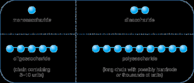
Carbohydrates are classified based on the number of sugar units:
Monosaccharides: Single sugar unit
Disaccharides: Two sugar units
Oligosaccharides: 3–10 sugar units
Polysaccharides: More than 10 sugar units
Monosaccharides
Monosaccharides are the simplest carbohydrates and cannot be hydrolyzed into smaller units. They are sweet, solid at room temperature, and highly soluble in water. Monosaccharides serve as building blocks for other carbohydrates.
Functional group: Aldoses (aldehyde group) or ketoses (ketone group)
Classification by carbon number: Trioses (3C), tetroses (4C), pentoses (5C), hexoses (6C), heptoses (7C)
Ketoses
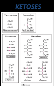
Ketoses are monosaccharides with a ketone functional group. Examples include dihydroxyacetone (3C), D-erythrulose (4C), D-ribulose and D-xylulose (5C), D-psicose, D-fructose, D-sorbose, and D-tagatose (6C).
Aldoses
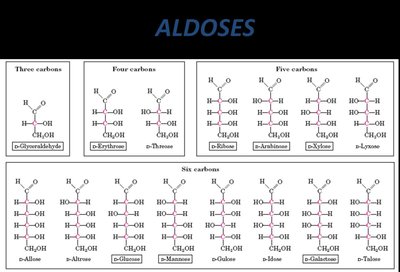
Aldoses are monosaccharides with an aldehyde functional group. Examples include D-glyceraldehyde (3C), D-erythrose and D-threose (4C), D-arabinose, D-ribose, D-xylose, D-lyxose (5C), and several hexoses (6C) such as D-glucose, D-mannose, D-galactose, D-talose.
Pentoses and Hexoses of Physiological Importance
Pentoses and hexoses play crucial roles in metabolism and cellular structure.
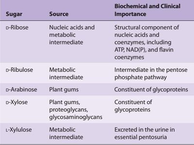
Pentoses
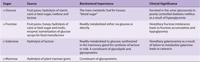
Sugar | Source | Biochemical and Clinical Importance |
|---|---|---|
D-Ribose | Nucleic acids and metabolic intermediate | Structural component of nucleic acids and coenzymes, including ATP, NAD(P), and flavin coenzymes |
D-Ribulose | Metabolic intermediate | Intermediate in the pentose phosphate pathway |
D-Arabinose | Plant gums | Constituent of glycoproteins |
D-Xylose | Plant gums, proteoglycans, glycosaminoglycans | Constituent of glycoproteins |
L-Xylulose | Metabolic intermediate | Excreted in the urine in essential pentosuria |
Hexoses
Sugar | Source | Biochemical Importance | Clinical Significance |
|---|---|---|---|
D-Glucose | Fruit juices, hydrolysis of starch, cane or beet sugar, maltose and lactose | Main metabolic fuel for tissues; "blood sugar" | Excreted in urine in diabetes mellitus as a result of hyperglycemia |
D-Fructose | Fruit juices, honey, hydrolysis of cane or beet sugar, hydrolysis of inulin, enzymatic isomerization of glucose syrup | Readily metabolized via glucose or directly | Hereditary fructose intolerance leads to fructose accumulation and hypoglycemia |
D-Galactose | Hydrolysis of lactose | Readily metabolized to glucose; constituent of glycolipids and glycoproteins | Hereditary galactosemia leads to cataracts |
D-Mannose | Hydrolysis of plant mannans/gums | Constituent of glycoproteins |
Stereochemistry of Monosaccharides
Chirality and Stereochemistry
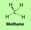
Monosaccharides possess one or more chiral (asymmetric) carbons, which are carbons attached to four different groups. The spatial arrangement of these groups determines the stereochemistry of the molecule. Most sugars used by humans are D-sugars.
Chiral carbon: Carbon atom with four different substituents
Representation: Wedges and dashed lines indicate bonds projecting in/out of the plane
Fischer Projection Formulas
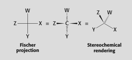
Fischer projections are a simplified way to represent the 3D structure of carbohydrates. The chiral carbon is at the intersection of vertical and horizontal lines. Horizontal bonds project out of the page, vertical bonds project into the page. The most oxidized carbon is placed at the top.
Isomerism in Monosaccharides
Types of Isomerism
Monosaccharides exhibit several types of isomerism:
Constitutional isomers: Same formula, different connectivity (e.g., glucose vs. fructose)
Stereoisomers: Same formula and connectivity, different spatial arrangement
Enantiomers: Mirror images, non-superimposable
Diastereomers: Not mirror images, non-superimposable
Epimers: Differ at only one chiral carbon
Anomers: Differ at the anomeric carbon formed during cyclization
Pyran-furan isomers: Ring size differences
Enantiomers
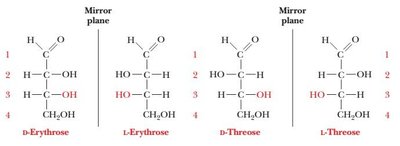
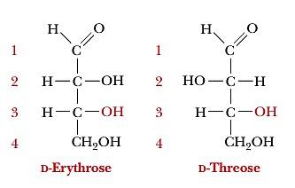
Enantiomers are stereoisomers that are mirror images of each other, such as D-erythrose and L-erythrose.
Diastereomers
Diastereomers are stereoisomers that are not mirror images, such as D-erythrose and D-threose.
Epimers
Epimers differ at only one chiral carbon. For example, D-glucose and D-mannose differ at C-2, while D-glucose and D-galactose differ at C-4.
Cyclization and Anomerism
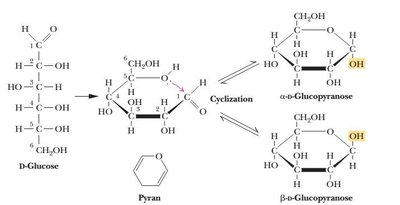
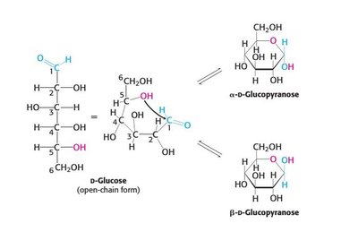
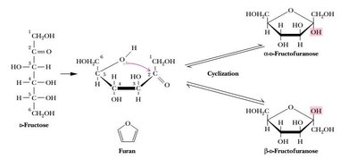
Monosaccharides with five or more carbons cyclize in solution, forming rings via intramolecular reactions between alcohol and aldehyde (hemiacetal) or ketone (hemiketal). The ring form is depicted using Haworth projections.
Anomerism
Cyclization creates a new chiral center (anomeric carbon), resulting in α and β anomers. The α-anomer has the hydroxyl group down, the β-anomer has it up. Anomers can interconvert via mutarotation.
Modified Monosaccharides
Sugar Acids
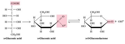
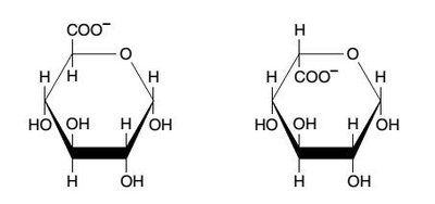
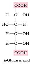
Sugar acids are formed by oxidation of monosaccharides, resulting in carboxyl groups. Types include:
Aldonic acids: Oxidation of the aldehyde group (e.g., gluconic acid)
Uronic acids: Oxidation of the terminal hydroxyl group (e.g., glucuronic acid)
Aldaric acids: Oxidation of both aldehyde and terminal hydroxyl (e.g., glucaric acid)
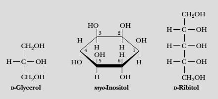
Sugar Alcohols
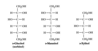
Sugar alcohols are produced by reduction of the carbonyl group in monosaccharides. They are sweet and used as sugar substitutes.
Alditols: Linear molecules, named by adding -itol (e.g., sorbitol, mannitol, xylitol)
Clinical relevance: Sorbitol accumulation in diabetics can cause cataracts
Biochemical roles: Ribitol in flavin coenzymes, glycerol and myo-inositol in lipids
Deoxy Sugars
Deoxy sugars have a hydroxyl group replaced by hydrogen. Examples include deoxyribose (component of DNA), L-fucose (component of glycoproteins), and L-rhamnose (component of ouabain).
Sugar Esters
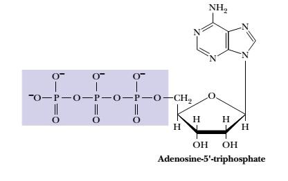
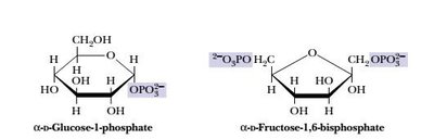
Phosphate esters of monosaccharides are important metabolic intermediates. Ribose in nucleotides (e.g., ATP, GTP) is phosphorylated at the 5' position.
Amino Sugars
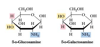
Amino sugars are hexoses where the hydroxyl group at carbon 2 is replaced by an amine group. Examples include D-glucosamine, D-galactosamine, and D-mannosamine.
Disaccharides
Structure and Examples
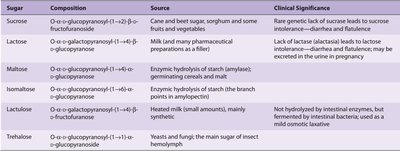
Disaccharides are formed by condensation of two monosaccharides via an O-glycosidic bond. Physiologically important disaccharides include maltose, sucrose, and lactose.
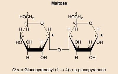
Maltose
Maltose consists of two α-D-glucose units joined by an α(1→4) glycosidic bond. Hydrolyzed by maltase.
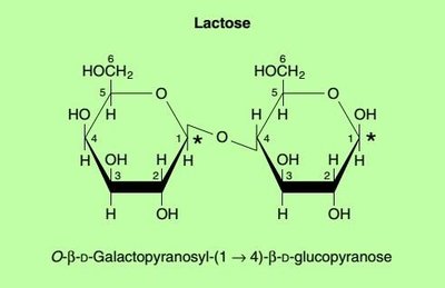
Lactose
Lactose is composed of β-D-galactose and α-D-glucose joined by a β(1→4) glycosidic bond. Hydrolyzed by lactase.
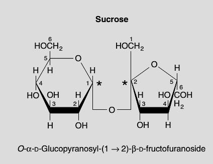
Sucrose
Sucrose is made of α-D-glucose and β-D-fructose joined by an α,β(1→2) glycosidic bond. Hydrolyzed by sucrase.
Cellobiose
Cellobiose is obtained from hydrolysis of cellulose and consists of two D-glucose units joined by β(1→4) linkages. Mammals cannot digest cellobiose.
Polysaccharides (Glycans)
Classification and Structure
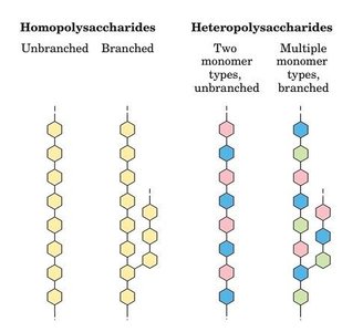
Polysaccharides are polymers of monosaccharides joined by O-glycosidic bonds. They are classified as homopolysaccharides (one type of monomer) or heteropolysaccharides (multiple types).
Starch
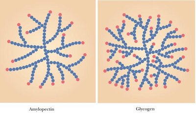
Starch is a homopolymer of glucose, serving as a storage carbohydrate in plants. It exists as amylose (linear, α(1→4) linkages) and amylopectin (branched, α(1→4) and α(1→6) linkages).
Glycogen
Glycogen is the major storage polysaccharide in animals, similar to amylopectin but more highly branched (branch points every 8–12 residues).
Inulin
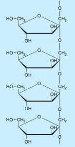
Inulin is a polysaccharide of fructose (fructosan) with β(2→1) linkages. Used to measure glomerular filtration rate.
Cellulose
Cellulose is a structural polysaccharide in plant cell walls, composed of β-D-glucose units with β(1→4) linkages. Mammals cannot digest cellulose.
Pectin
Pectin is a heteropolysaccharide mainly of β-D-galacturonic acid, used as a gelling agent in foods.
Chitin
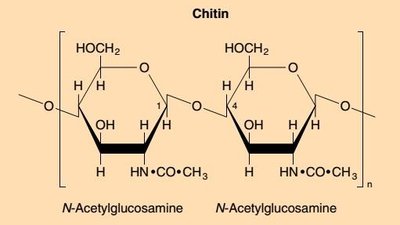
Chitin is a structural polysaccharide in exoskeletons and cell walls, composed of N-acetylglucosamine units with β-glycosidic bonds.
Agarose and Agar
Agarose is a linear polymer of agarobiose (D-galactose and 3,6-anhydro-L-galactose), used in electrophoresis. Agar is a mixture of agarose and agaropectin, used as a culture medium and thickener.
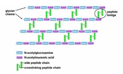
Peptidoglycan (Murein)
Peptidoglycan is a structural polysaccharide in bacterial cell walls, composed of N-acetylglucosamine and N-acetylmuramic acid with β(1→4) linkages, cross-linked by peptides.
Glycosaminoglycans (GAGs)
Structure and Classification
GAGs are complex heteropolysaccharides found in the extracellular matrix, composed of repeating disaccharide units of acetylated amino sugars and uronic acids. Some are sulfated.
Classes: Hyaluronic acid, heparin, heparan sulfate, keratan sulfate, dermatan sulfate, chondroitin-4-sulfate, chondroitin-6-sulfate
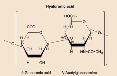
Hyaluronic Acid
Contains D-glucuronic acid and N-acetylglucosamine. Functions as a lubricant and shock absorber in joints, and is a component of the vitreous humor and extracellular matrix.
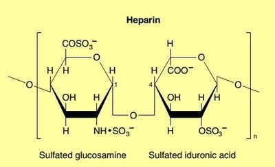
Heparin
Intracellular GAG with high negative charge density. Natural anticoagulant, binds antithrombin.
Heparan Sulfate
Similar to heparin but with fewer sulfate groups and some acetylated amino groups. Extracellular GAG.
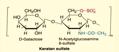
Keratan Sulfate
Contains D-galactose and N-acetylglucosamine. Only GAG without uronic acid. Found in cornea, cartilage, tendons, bones, and "horny" structures.
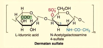
Dermatan Sulfate
Contains L-iduronic acid and N-acetylgalactosamine. Contributes to skin pliability and is present in blood vessels, heart valves, and sclera of the eye.
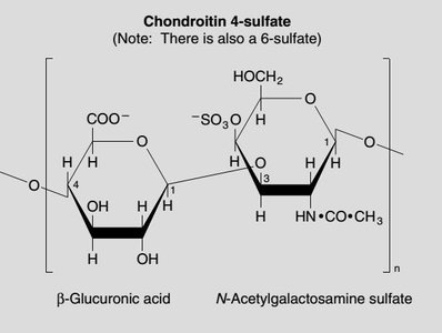
Chondroitin Sulfates
Contains D-glucuronic acid and N-acetylgalactosamine sulfate. Most abundant GAG, found in cartilage, tendons, ligaments, and aorta walls.
Glycoconjugates
Types and Functions
Glycoconjugates are compounds where carbohydrates are covalently linked to other macromolecules. Types include proteoglycans, glycoproteins, glycolipids, and lipopolysaccharides.
Proteoglycans
Carbohydrates (mainly GAGs) covalently linked to proteins, found on cell surfaces and in the extracellular matrix. The carbohydrate portion is the main site of biological activity.
Glycoproteins
Proteins with covalently attached carbohydrates (mono-, di-, or oligosaccharides). The protein content is higher than the carbohydrate content.
Classification of Glycoproteins
O-linked: Carbohydrate attached to hydroxyl side chains of serine or threonine
N-linked: Carbohydrate attached to amide nitrogen of asparagine
GPI-linked: Linked to carboxyl terminal amino acid via phosphoryl-ethanolamine moiety joined to an oligosaccharide, linked via glucosamine to phosphatidylinositol
Functions of Glycoproteins
Structural molecules (e.g., collagen)
Cell surface receptors for hormones and drugs
Hormones (e.g., FSH, TSH, hCG)
ABO blood group substances
Enzymes (e.g., alkaline phosphatase)
Pathogen attachment (e.g., HIV, Influenza A)
