 뒤로
뒤로The Structure and Function of Large Biological Molecules: Foundations of Biochemistry
스터디 가이드 - 스마트 노트
 자료에 맞춘 맞춤형 노트, 핵심 정의, 예시, 맥락을 확장해 제공합니다.
Introduction to Organic Chemistry and Biochemistry
Vitalism vs. Mechanism
Early scientific thought distinguished between vitalism—the belief that organic compounds could only arise within living organisms—and mechanism, which posited that all natural phenomena are governed by physical and chemical laws. The Stanley Miller Experiment provided experimental evidence supporting mechanism by demonstrating that organic molecules could be synthesized abiotically under conditions thought to resemble those of early Earth.
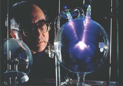
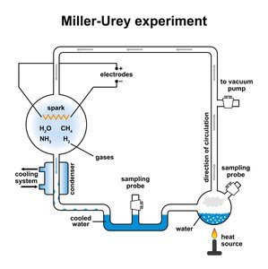
Key Point: The Miller-Urey experiment simulated early Earth conditions and produced amino acids and other organic molecules, suggesting that life's building blocks could form naturally.
Example: 13 of the 20 amino acids, nitrogenous bases, and adenine were synthesized in the experiment.
Carbon: The Backbone of Biological Molecules
Properties of Carbon
Carbon is central to organic chemistry due to its tetravalence (four valence electrons), allowing it to form four covalent bonds and a variety of stable structures. The geometry of carbon's bonds is tetrahedral, and the diversity of carbon skeletons (length, branching, rings, double/triple bonds) underlies the diversity of organic molecules.
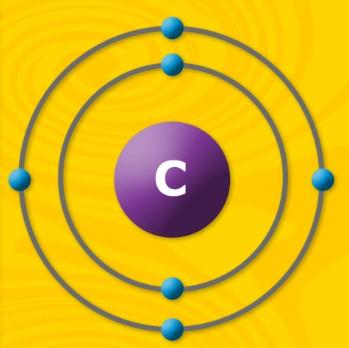
Key Point: Carbon's ability to form four bonds enables the construction of complex molecules essential for life.
Example: Hydrocarbons, carbohydrates, proteins, lipids, and nucleic acids all have carbon backbones.
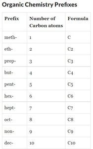
Nomenclature and Types of Hydrocarbons
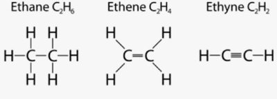
Hydrocarbons are compounds composed only of carbon and hydrogen. They are classified based on the types of bonds between carbon atoms:
Alkanes: Only single bonds (saturated); general formula ending in (-ane).
Alkenes: At least one double bond (unsaturated); general formula ending in (-ene).
Alkynes: At least one triple bond (unsaturated); general formula ending in (-yne).
Cycloalkanes: Have one or more rings of carbons
Isomerism in Organic Molecules
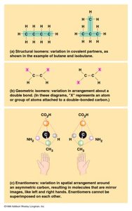
Isomers are molecules with the same molecular formula but different structures and properties -> structural & stereoisomers
Structural : Differ in covalent bonding arrangement of atoms.
Geometric (cis-trans) isomers: Differ in spatial arrangement around a double bond (C=C).
Enantiomers: Mirror images due to an asymmetric carbon; important in pharmacology (e.g., thalidomide).
Functional Groups in Organic Molecules
Overview of Functional Groups
Functional groups are specific groups of atoms attached to carbon skeletons that give organic molecules their unique behaviors and properties. They are critical in determining the behavior and reactivity of biomolecules. An alkane that has lost a H so it can bond to a carbon is called an alkyl (group).
Hydroxyl (-OH): Found in alcohols; polar and soluble in water (hydrogen bonds).
Carbonyl (C=O): Found in aldehydes (end of chain) and ketones (within chain).
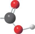
Carboxyl (-COOH): Found in carboxylic acids; acts as an acid (can donate H+).
Amino (-NH2): Found in amines; acts as a base (can accept H+).
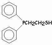
Sulfhydryl (-SH): Found in thiols; can form disulfide bonds in protein to stabilize structure.
Phosphate (-PO4): Found in organic phosphates; important in energy transfer (e.g., ATP).
Macromolecules: Structure and Function
Polymers and Monomers
Most biological macromolecules are polymers, long chains of repeating units (covalently bonded) called monomers. The four major classes are carbohydrates, proteins, nucleic acids, and lipids (not true polymers).
Polymerization: Monomers are joined by dehydration (condensation) reactions, releasing water. Polymers are broken down by hydrolysis, which adds water to break bonds.
Dehydration synthesis releases water (removes a water molecule & forming a new bond) to build larger molecules, while hydrolysis adds water (a water molecule & breaking a bond) to break them apart.
Carbohydrates
Monosaccharides
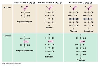
Monosaccharides are simple sugars with the general formula . They contain multiple hydroxyl (-OH) groups and one carbonyl (C=O) group, which can be at the end (aldose) or within the chain (ketose).
Examples: Glucose (an aldose), fructose (a ketose), ribose (pentose sugar in RNA).
Disaccharides and Glycosidic Linkages
Disaccharides are formed by joining two monosaccharides via a glycosidic linkage (covalent bond), typically through a dehydration reaction.
Examples: Sucrose (glucose + fructose), maltose (glucose + glucose), lactose (glucose + galactose).
Polysaccharides: Storage and Structure
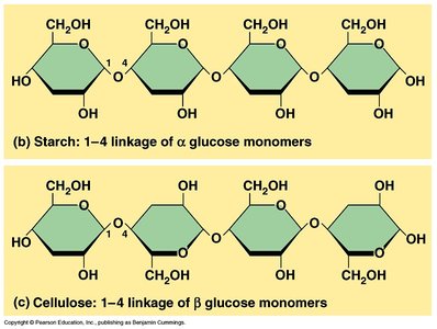
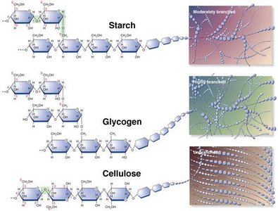
Polysaccharides are long chains of monosaccharides (Glucose molecules). Their function depends on the type of monomer and the type of glycosidic linkage.
Storage: Starch (plants), glycogen (animals)
Structure: Cellulose (plants), chitin (fungi, arthropods)
Alpha (α) vs. Beta (β) Glucose and Linkages
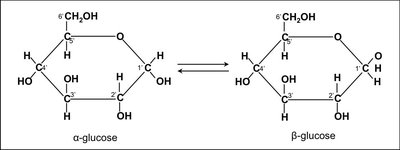
The orientation of the hydroxyl group on the anomeric carbon (C1) distinguishes α-glucose from β-glucose, leading to different polymer structures and digestibility.
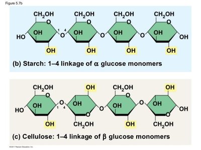
Alpha (α) glucose: OH on C1 is down; forms helical polymers (e.g., starch, glycogen).
Beta (β) glucose: OH on C1 is up; forms straight chains/linear (e.g., cellulose).
Enzymatic specificity: Humans can digest α-linkages but not β-linkages (cellulose is dietary fiber). We can digest things like starch & glycogen in which our enzymes can break down easily. Our enzymes can't get into beta type linkages because there is not enough space, so it's not broken down easily.
Lipids
Structure and Types of Lipids
Lipids are hydrophobic molecules, not true polymers, composed mainly of hydrocarbons. Major types include fats, phospholipids, and steroids.
Fats (triglycerides): Glycerol + 3 fatty acids, joined by ester linkages (dehydration formation)
Saturated fats: Only single bonds; solid at room temperature, easy to stack.
Unsaturated fats: One or more double bonds; liquid at room temperature, bend.
Phospholipids: Glycerol + 2 fatty acids + phosphate group; form cell membranes (bilayers), with hydrophobic tail & hydrophilic head.
Steroids: Four fused carbon rings (e.g., cholesterol, sex hormones).
Proteins
Amino Acids and Peptide Bonds
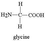
Proteins are polymers of amino acids, each containing an amino group (NH2), carboxyl group (-COOH), hydrogen (H), and variable R group. Polypeptides : Peptide bonds (covalent bond) link amino acids via dehydration reactions. Proteins nearly have limitless combinations -> 20^n (n = how long is the amino acids)
Primary structure: Linear sequence of amino acids.
Secondary structure: Alpha helices and beta sheets formed by hydrogen bonding.
Tertiary structure: A single polypeptide chain. 3D folding due to R group interactions (hydrophobic, ionic, hydrogen bonds. disulfide bridges : a strong covalent bond that stabilizes protein).
Quaternary structure: Multiple polypeptide chains assembled together into 1 macromolecule.
3D shape of a protein is critical to its function. Anything that changes (denatures) the shape of a protein can alter or destroy its function. Any change in any structure can change the other structures and leaving it useless & possibly cause genetic disease. Proteins denatures when temperatures get to high or pH gets to extreme.
Enzymtic : Special kinds of proteins in living things that can help break molecules or build molecules.
Structural : Proteins for support.
Storage : Proteins functioning for storage of amino acids.
Transport : Proteins for transport of other substances.
Hormonal : Proteins for the coordination of an organism' activities.
Receptor : Proteins for response of cell to chemical stimuli.
Contractile & Motor : Proteins for movement.
Defensive : Proteins for protection against disease.
Nucleic Acids
DNA and RNA Structure
Nucleic acids are polymers of nucleotides, each consisting of a nitrogenous base, pentose sugar, and phosphate group. DNA and RNA differ in their sugars (deoxyribose vs. ribose) and bases (thymine in DNA, uracil in RNA).
Base pairing: Purines (adenine, guanine) pair with pyrimidines (thymine/uracil, cytosine).
Purines have double rings, pyrimidines have single rings.
Pentose : Ribose (RNA), deoxyribose (DNA), nucleoside (base + sugar)
Phosphodiester bonds/linkages: Link nucleotides (covalent bond between phosphate + sugar) in the backbone.
Double helix: DNA strands run antiparallel (5' to 3') -> The two strands of a DNA molecule run side by side, but they point in opposite directions.
Strand One runs downward (5->3).
Strand Two runs upward right next to it (3->5).
Inheritance based on DNA replication.
According to Chargaff's Rule : If we know 1 amount of the 4 N bases, we can calculate the other 3 bases' amounts.
Ex : DNA has 15% cytosine,then Guanine also has 15% since C=G.
Ex : Accounts for 30%, A & T make up 70% total, with 35& each since A=T.
Summary Table: Macromolecules
Macromolecule | Monomer | Polymer | Bond Type | Examples |
|---|---|---|---|---|
Carbohydrates | Monosaccharides | Disaccharides/Polysaccharides | Glycosidic Linkages (α and β) | Glucose, starch |
Proteins | Amino Acids | Polypeptide (protein) | Peptide (H-bonds, ionic, disulfide bridges) | Enzymes, antibodies, muscle |
Lipids | Fatty Acids | (Not a true polymer) | Ester linkages | Phospholipids, triglycerides, steroids |
Nucleic Acids | Nucleotide | DNA or RNA | Phosphodiester linkages (also H-bonds, Van der Waals) | DNA/RNA |
