 뒤로
뒤로A Tour of the Cell: Structure and Function of Cells
스터디 가이드 - 스마트 노트
 자료에 맞춘 맞춤형 노트, 핵심 정의, 예시, 맥락을 확장해 제공합니다.
Chapter 6: A Tour of the Cell
Introduction to Cells
Cells are the fundamental structural and functional units of all living organisms. Organisms may be unicellular (composed of a single cell, such as bacteria) or multicellular (composed of many cells, such as plants and animals). Understanding cell structure and function is essential for all biological sciences.
Types of Cells
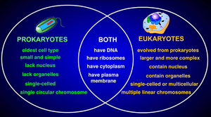
Prokaryotic Cells: Simple, small cells lacking a true nucleus and membrane-bound organelles. Their genetic material is found in the cytoplasm. Examples include bacteria and archaea.
Eukaryotic Cells: Larger, more complex cells with a membrane-bound nucleus and specialized organelles. Examples include animal, plant, fungal, and protist cells.
Internal Organization of Eukaryotic Cells
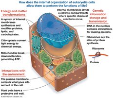
Eukaryotic cells contain internal membranes that divide the cell into compartments called organelles. These compartments allow for specialized functions and the separation of incompatible biochemical processes.
6.1: Studying Cells
Microscopy
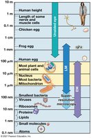
Cells are typically too small to be seen with the naked eye. Microscopes are essential tools for visualizing cells and their components. There are several types of microscopy, each with unique advantages:
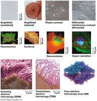
Light Microscopy (LM): Uses visible light and glass lenses to magnify images up to about 1,000 times. Useful for viewing live cells and general cell structure.
Electron Microscopy (EM): Uses electron beams for much higher resolution. Includes:
Scanning Electron Microscopy (SEM): Provides 3D images of cell surfaces.
Transmission Electron Microscopy (TEM): Reveals internal cell structures.
Fluorescence and Confocal Microscopy: Use fluorescent markers and optical sectioning for detailed imaging of specific cell components.
Cryo-Electron Microscopy: Preserves specimens at very low temperatures, allowing visualization of structures in their native state.
Cell Fractionation
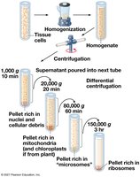
Cell fractionation is a laboratory technique used to separate cellular components for individual study. This is typically achieved by homogenizing cells and then using differential centrifugation to separate organelles based on size and density.
Comparing Prokaryotic and Eukaryotic Cells
Basic Features of All Cells
Plasma membrane: Selective barrier that encloses the cell.
Cytosol: Semifluid substance within the cell.
Chromosomes: Carry genetic information (DNA).
Ribosomes: Sites of protein synthesis.
Key Differences
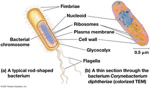
Prokaryotic Cells: DNA is located in a region called the nucleoid, not enclosed by a membrane. Lack membrane-bound organelles.
Eukaryotic Cells: DNA is enclosed within a double-membrane nucleus. Contain various membrane-bound organelles.
Size: Eukaryotic cells are generally much larger than prokaryotic cells.
Surface Area to Volume Ratio
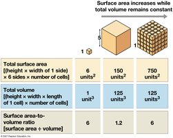
The surface area-to-volume ratio is critical for cell function. As a cell grows, its volume increases faster than its surface area, limiting the rate of material exchange with the environment.
A Panoramic View of the Eukaryotic Cell
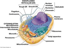
Eukaryotic cells have a variety of organelles, each with specialized functions. Internal membranes create compartments that allow for diverse cellular processes.
6.3: The Nucleus and Ribosomes
The Nucleus: Information Central
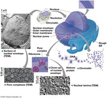
The nucleus is the control center of the cell, containing most of the cell's genetic material. It is surrounded by a double membrane called the nuclear envelope, which contains nuclear pores for molecular transport.
Chromatin: DNA-protein complex that condenses to form chromosomes during cell division.
Nucleolus: Site of ribosomal RNA (rRNA) synthesis and ribosome assembly.
Nuclear Lamina: Protein network that maintains nuclear shape.
Ribosomes: Protein Factories
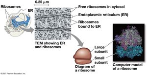
Ribosomes are complexes of rRNA and protein that synthesize proteins. They can be found free in the cytosol or bound to the endoplasmic reticulum (ER) or nuclear envelope.
6.4: The Endomembrane System
Components and Functions
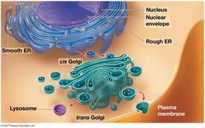
The endomembrane system is a network of membranes involved in protein and lipid synthesis, modification, and transport. It includes the nuclear envelope, endoplasmic reticulum (ER), Golgi apparatus, lysosomes, vacuoles, and plasma membrane.
Endoplasmic Reticulum (ER)
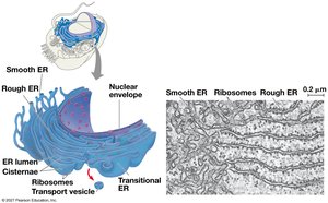
Smooth ER: Lacks ribosomes; synthesizes lipids, detoxifies drugs, and stores calcium ions.
Rough ER: Studded with ribosomes; synthesizes proteins and membranes, distributes transport vesicles.
Golgi Apparatus
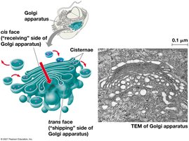
The Golgi apparatus modifies, sorts, and packages proteins and lipids for storage or transport out of the cell. It consists of flattened sacs called cisternae.
Lysosomes
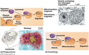
Lysosomes are membrane-bound sacs containing hydrolytic enzymes for digesting macromolecules. They are involved in phagocytosis (ingestion of external particles) and autophagy (recycling of cellular components).
Vacuoles
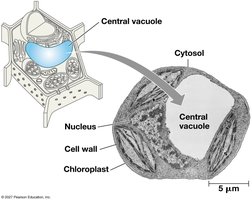
Vacuoles are large vesicles with diverse functions. Plant cells have a central vacuole that stores ions and contributes to cell growth. Contractile vacuoles in protists expel excess water.
Endomembrane System Overview
The endomembrane system coordinates the synthesis, modification, and transport of cellular products.
6.5: Mitochondria and Chloroplasts
Endosymbiont Theory
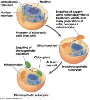
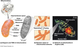
Mitochondria and chloroplasts are energy-converting organelles. The endosymbiont theory proposes that these organelles originated as free-living bacteria engulfed by ancestral eukaryotic cells. Evidence includes their double membranes, own DNA, and ribosomes.
Mitochondria: Cellular Respiration
Mitochondria are the sites of cellular respiration, generating ATP from organic molecules using oxygen. They have an outer membrane and a highly folded inner membrane (cristae), creating compartments for metabolic reactions.
Chloroplasts: Photosynthesis
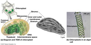
Chloroplasts are found in plants and algae and are the sites of photosynthesis. They contain thylakoids (membranous sacs) stacked into grana and surrounded by stroma (fluid).
Peroxisomes
Peroxisomes are single-membrane organelles containing enzymes that transfer hydrogen from substrates to oxygen, producing hydrogen peroxide. They are involved in fatty acid breakdown and detoxification.
6.6: The Cytoskeleton
Structure and Function
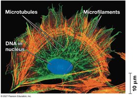
The cytoskeleton is a network of protein fibers that provides structural support, organizes cell components, and enables cell movement. It consists of three main types of fibers:
Microtubules: Hollow rods made of tubulin; involved in cell shape, organelle movement, and chromosome separation.
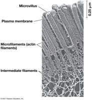
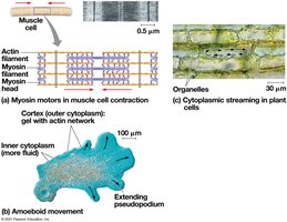
Microfilaments (Actin Filaments): Thin rods of actin; support cell shape and are involved in cell movement and muscle contraction.
Intermediate Filaments: Fibrous proteins; provide mechanical support and anchor organelles.
Motor Proteins and Cell Motility
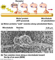
Motor proteins interact with cytoskeletal elements to produce movement of organelles and vesicles within cells.
Centrosomes and Centrioles
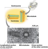
In animal cells, microtubules originate from the centrosome, which contains a pair of centrioles. These structures are important for organizing microtubules during cell division.
Cilia and Flagella
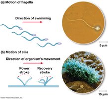
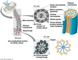
Cilia and flagella are microtubule-containing extensions that enable cell movement. Cilia are usually numerous and short, while flagella are longer and fewer. Both have a "9+2" arrangement of microtubules and are powered by the motor protein dynein.
Microfilaments and Cell Movement
Microfilaments support cell shape and are involved in movements such as muscle contraction, amoeboid movement, and cytoplasmic streaming in plant cells.
6.7: Extracellular Components and Cell Junctions
Cell Walls of Plants
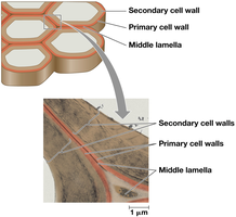
Plant cells are surrounded by a rigid cell wall made of cellulose, which provides structural support, protection, and prevents excessive water uptake. Cell walls may have multiple layers: primary cell wall, middle lamella, and secondary cell wall.
Extracellular Matrix (ECM) of Animal Cells
Animal cells lack cell walls but are surrounded by an extracellular matrix (ECM) composed of glycoproteins like collagen, proteoglycans, and fibronectin. The ECM provides structural support and mediates cell signaling.
Cell Junctions
Plasmodesmata: Channels between plant cells that allow transport of water, ions, and small molecules.
Tight Junctions: Seal neighboring animal cells together to prevent leakage of extracellular fluid.
Desmosomes: Anchor animal cells together into strong sheets.
Gap Junctions: Provide cytoplasmic channels for communication between adjacent animal cells.
6.8: Integration of Cellular Components
The cell functions as an integrated unit, with all components working together to maintain life. For example, the destruction of bacteria by a macrophage involves coordination between the cytoskeleton, lysosomes, and plasma membrane.
