 뒤로
뒤로A Tour of the Cell: Structure, Function, and Diversity
스터디 가이드 - 스마트 노트
 자료에 맞춘 맞춤형 노트, 핵심 정의, 예시, 맥락을 확장해 제공합니다.
A Tour of the Cell
Cell Theory
The cell theory is a fundamental concept in biology, describing the basic properties of cells and their role in life.
All organisms consist of one or more cells: Every living thing is composed of cells, whether unicellular or multicellular.
The cell is the simplest collection of matter that can live: Cells are the smallest units capable of performing all life functions.
All cells arise only from preexisting cells: New cells are produced by the division of existing cells.
Cell structure is related to cellular function: The shape and internal organization of a cell are closely linked to its role in the organism.
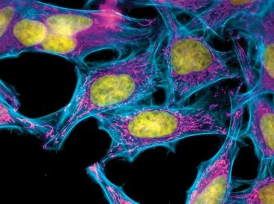
Microscopy and Cell Visualization
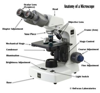
Microscopes are essential tools for studying cells, as most are too small to be seen with the naked eye.
Compound Light Microscope (LM): Uses visible light and glass lenses to magnify specimens up to 1000x.
Magnification: The increase in apparent size of an object.
Resolution: The clarity of an image; the ability to distinguish two close objects as separate.
Electron Microscopes (EM): Use electron beams for much higher resolution (up to 1,000,000x), including Scanning (SEM) and Transmission (TEM) types.
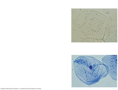
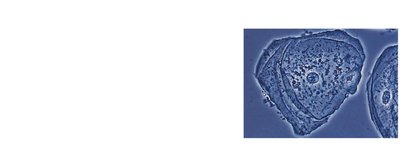
Staining and Contrast Techniques: Stains, dyes, phase-contrast, fluorescence, and confocal microscopy enhance visibility and detail of cell structures.
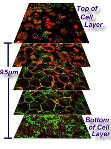
Cell Size and Surface-to-Volume Ratio
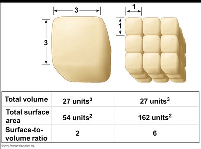
Cell size is limited by the need to efficiently exchange materials with the environment. The surface-to-volume ratio is critical for this exchange.
Cells must be large enough to contain DNA, proteins, and organelles, but small enough for efficient material exchange.
Surface-to-volume ratio: Smaller cells have a higher ratio, allowing more efficient exchange.
Total volume | Total surface area | Surface-to-volume ratio |
|---|---|---|
27 units3 | 54 units2 | 2 |
27 units3 | 162 units2 | 6 |
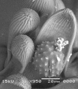
Types of Cells: Prokaryotic vs. Eukaryotic
Cells are classified as prokaryotic or eukaryotic based on their internal structure.
Prokaryotic cells: Found in domains Bacteria and Archaea; lack a nucleus and membrane-bound organelles; DNA is in the nucleoid region.
Eukaryotic cells: Found in protists, fungi, animals, and plants; have a nucleus and membrane-bound organelles; DNA is enclosed within the nucleus.
Common features: Both types have a plasma membrane, cytoplasm, chromosomes (DNA), and ribosomes.

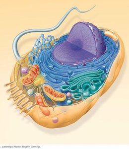
Functional Compartments of Eukaryotic Cells
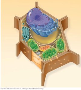
Eukaryotic cells are organized into compartments, each with specialized functions.
Genetic control: Nucleus and ribosomes
Manufacture, distribution, and breakdown: Endoplasmic reticulum, Golgi apparatus, lysosomes, vacuoles
Energy processing: Mitochondria (all cells), chloroplasts (plants and some protists)
Structural support, movement, communication: Cytoskeleton, plasma membrane, cell wall
The Plasma Membrane
The plasma membrane is a selective barrier that controls the movement of substances in and out of the cell.
Structure: Double layer of phospholipids with embedded proteins.
Function: Maintains homeostasis by regulating transport.
The Nucleus and Chromosomes
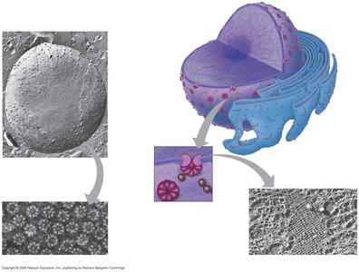
The nucleus is the genetic control center of the cell, containing most of the cell's DNA.
Nuclear envelope: Double membrane with pores for molecular transport.
Chromatin: DNA and proteins; condenses to form chromosomes during cell division.
Nucleolus: Site of ribosome assembly.

Ribosomes: Protein Factories
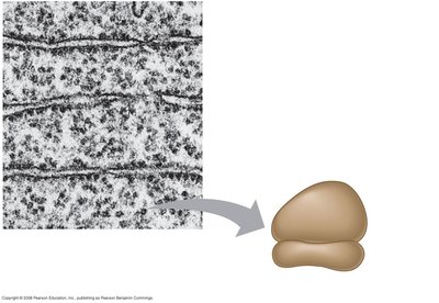
Ribosomes are responsible for protein synthesis, using instructions from DNA.
Structure: Made of ribosomal RNA (rRNA) and proteins; consists of large and small subunits.
Types: Free ribosomes (in cytoplasm) and bound ribosomes (attached to ER or nuclear envelope).
The Endomembrane System
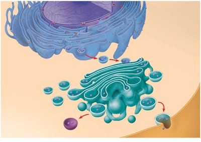
The endomembrane system is a network of membranes involved in synthesis, transport, and breakdown of molecules.
Components: Nuclear envelope, endoplasmic reticulum (ER), Golgi apparatus, lysosomes, vacuoles, plasma membrane, vesicles.
Function: Coordinates cellular activities and molecular trafficking.
Endoplasmic Reticulum (ER)
Rough ER: Studded with ribosomes; synthesizes glycoproteins, distributes transport vesicles, and produces membrane components.
Smooth ER: Lacks ribosomes; synthesizes lipids, hydrolyzes carbohydrates, detoxifies poisons, stores calcium.
Golgi Apparatus
Structure: Flattened membranous sacs (cisternae).
Function: Modifies, sorts, and packages products from the ER; produces lysosomes.
Lysosomes
Structure: Membrane-bound sacs of hydrolytic enzymes.
Function: Digests macromolecules, recycles cell components (autophagy), breaks down ingested material (phagocytosis).
Vacuoles
Structure: Large vesicles with diverse functions.
Function: Store water and organic compounds, hydrolyze materials, maintain cell structure (especially in plants).
Mitochondria and Chloroplasts: Energy Organelles
Mitochondria and chloroplasts are responsible for energy transformation in cells.
Mitochondria: Sites of cellular respiration; use oxygen to generate ATP from chemical energy; have double membranes, their own DNA and ribosomes, and replicate independently.
Chloroplasts: Sites of photosynthesis in plants and some protists; convert solar energy to chemical energy; contain their own DNA and ribosomes.
Endosymbiotic Theory
The endosymbiotic theory explains the origin of mitochondria and chloroplasts as formerly independent prokaryotes that were engulfed by ancestral eukaryotic cells.
Evidence: Both organelles have double membranes, their own circular DNA, ribosomes, and replicate independently.
The Cytoskeleton
The cytoskeleton is a network of fibers that organizes cell structure, movement, and communication.
Types: Microtubules, microfilaments, intermediate filaments.
Functions: Support cell shape, anchor organelles, facilitate movement, regulate biochemical activities.
Centrosomes and Centrioles
Centrosome: Microtubule-organizing center in animal cells; contains a pair of centrioles.
Centrioles: Each has nine triplets of microtubules arranged in a ring.
Cilia and Flagella
Structure: Both have a "9+2" arrangement of microtubules.
Function: Cilia move substances across cell surfaces; flagella propel cells.
Extracellular Structures and Intercellular Junctions
Cells synthesize and secrete materials for support and communication.
Cell walls: Found in plants, fungi, and some protists; made of cellulose, polysaccharides, and protein; provide protection and shape.
Extracellular matrix (ECM): In animal cells; composed of glycoproteins like collagen; functions in support, adhesion, movement, and regulation.
Intercellular junctions: Facilitate cell adhesion and communication; types include plasmodesmata (plants), tight junctions, desmosomes, and gap junctions (animals).
Summary Table: Comparison of Cell Types
Cell Type | Nucleus | Membrane-bound Organelles | Cell Wall | Chloroplasts | Mitochondria |
|---|---|---|---|---|---|
Prokaryotic (Bacteria) | No | No | Yes | No | No |
Fungal | Yes | Yes | Yes | No | Yes |
Protist | Yes | Yes | Some | Some | Yes |
Animal | Yes | Yes | No | No | Yes |
Plant | Yes | Yes | Yes | Yes | Yes |
Additional info: These notes expand on brief points from the original materials, providing definitions, examples, and context for each topic. All images included are directly relevant to the adjacent explanations, reinforcing key concepts in cell biology.
