 뒤로
Cancer and Cell Division: Mitosis, Cell Cycle Regulation, and Genetic Mutations
스터디 가이드 - 스마트 노트
 자료에 맞춘 맞춤형 노트, 핵심 정의, 예시, 맥락을 확장해 제공합니다.
Cancer and Cell Division
Introduction to Cancer and the Cell Cycle
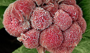
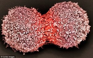
Cancer is fundamentally a disease of the cell cycle, arising when mutations accumulate in genes that regulate cell division. These mutations disrupt normal cell cycle control, leading to uncontrolled cell proliferation.
Tumor suppressor genes: Genes such as p53 and BRCA1/BRCA2 that pause the cell cycle or initiate apoptosis.
Proto-oncogenes: Genes like ras that promote cell cycle progression; when mutated, they become oncogenes and drive excessive cell division.
DNA repair genes: Maintain genome integrity by repairing DNA damage.
Mutations and Their Sources
Mutations are changes in the DNA sequence and are the primary source of genetic errors leading to cancer.
DNA replication errors: Mistakes during DNA synthesis.
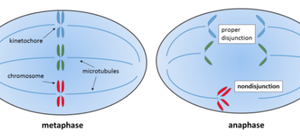
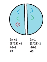
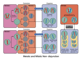
Non-disjunction: Failure of chromosomes to separate properly during mitosis or meiosis.
Exposure to mutagens: Environmental agents such as radiation or chemicals that damage DNA.
Cell Division: Mitosis and Meiosis
Why Do Cells Divide?
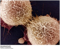
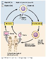
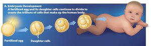
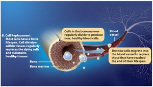
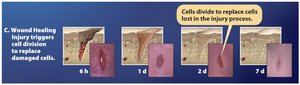
Cell division is essential for growth, development, cell replacement, and wound healing.
Growth and development: Formation of multicellular organisms from a single cell.
Cell replacement: Replacement of dying cells to maintain tissue health.
Wound healing: Repair of damaged tissues.
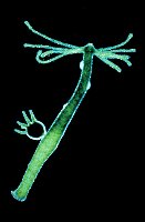
Asexual reproduction: Some organisms reproduce by mitosis (e.g., hydra).
Mechanisms of Cell Division
Cells do not simply split in half; they must distribute a complete set of genetic material to each daughter cell.
Genome size: Varies among organisms (e.g., prokaryotes: 2 million bp, humans: 3 billion bp).
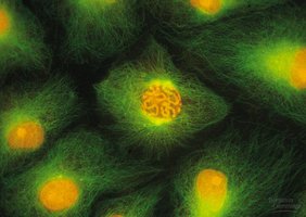
Eukaryotic chromosomes: Highly organized structures that ensure accurate distribution of genetic material.
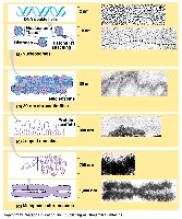
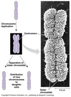
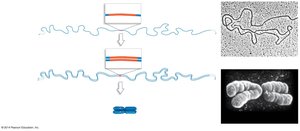
Karyotype Preparation
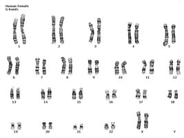
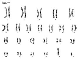
Karyotyping is used to visualize chromosomes and detect abnormalities.
G-banding: Staining technique to identify individual chromosomes.
Human karyotype: Shows the complete set of chromosomes in a cell.
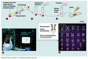
The Cell Cycle
Phases of the Cell Cycle
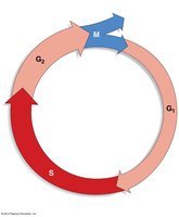
The cell cycle consists of two main phases:
Interphase: Includes G1 (first gap), S (DNA synthesis), and G2 (second gap) phases. Cells grow, replicate DNA, and prepare for division.
M phase (mitosis): Nuclear division followed by cytokinesis.
Interphase Details
S phase: DNA replication occurs.
G1 and G2 phases: Cell growth, organelle replication, and preparation for division.
Duration: Typical cell cycle lasts about 24 hours.
Mitosis: Steps and Mechanisms
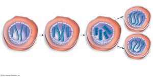
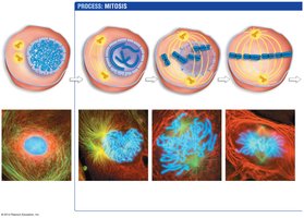
Mitosis is divided into several subphases:
Prophase: Chromosomes condense, spindle apparatus forms.
Prometaphase: Nuclear envelope breaks down, microtubules attach to kinetochores.
Metaphase: Chromosomes align at the cell's equator.
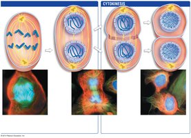
Anaphase: Sister chromatids separate and move to opposite poles.
Telophase: Nuclear envelope reforms, chromosomes decondense.
Cytokinesis: Division of cytoplasm and organelles.
Cytokinesis in Different Cell Types
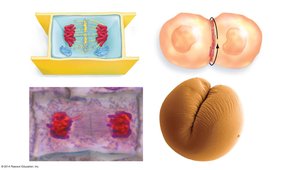
Plants: Vesicles form a cell plate.
Animals, fungi, slime molds: Actin-myosin ring forms a cleavage furrow.
Bacteria: Divide by binary fission, not cytokinesis.
Control of the Cell Cycle
Internal and External Controls
Cell cycle regulation is achieved through internal and external mechanisms.
Internal controls: Cyclins, protein kinases, proto-oncogenes, tumor suppressors.
External controls: Growth factors, hormones, density-dependent inhibition, anchorage dependence.
Cell-Cycle Checkpoints
There are three main checkpoints:
G1 checkpoint: Ensures cell size, nutrients, and DNA integrity.
G2 checkpoint: Ensures DNA replication is complete and undamaged.
M-phase checkpoint: Ensures chromosomes are properly attached and segregated.
Cancer: Out-of-Control Cell Division
Genetic Basis of Cancer
Cancer arises from mutations in genes that regulate cell division and DNA repair.
Multi-hit hypothesis: Multiple mutations are required for cancer development.
Oncogenes: Mutated proto-oncogenes that promote cell division.
Tumor suppressor genes: Loss of function leads to uncontrolled division.
DNA repair genes: Defects increase mutation rates.
Properties of Cancer Cells
Malignant tumors: Invasive, can metastasize (spread to other tissues).
Benign tumors: Non-invasive, do not spread.
Key Genes in Cancer
Proto-oncogenes (e.g., Ras): Promote cell division; mutations lead to constant activation.
Tumor suppressors (e.g., p53, BRCA1/BRCA2): Inhibit cell division or repair DNA; mutations inactivate these functions.
Apoptosis genes: Regulate programmed cell death.
DNA repair genes: Maintain genome stability.
Cell Cycle Checkpoints and Cancer
p53 protein: Pauses cell cycle, activates DNA repair, or initiates apoptosis if damage is irreparable.
BRCA1/BRCA2: Involved in DNA repair, especially double-stranded breaks.
External Control and Cancer Therapy
External Controls and Cancer Cell Behavior
Normal cells: Exhibit anchorage dependence and density-dependent inhibition.
Cancer cells: Escape these controls, leading to uncontrolled growth.
Cancer Treatments
Chemotherapy: Uses drugs to interfere with cell division (e.g., spindle formation, DNA replication).
Radiation therapy: Uses high-energy beams to damage DNA and trigger apoptosis.
Immunotherapies: New approaches targeting cancer cells via the immune system.
Prevention and Lifestyle
Mutagens: Cancer-causing agents (e.g., X-rays, UV radiation, tobacco).
Healthy lifestyle: Avoiding carcinogens, eating fiber and antioxidants, regular checkups.
Summary Table: Cell Cycle and Cancer
Comparison of Key Genes and Their Roles
Gene Type | Normal Function | Mutation Effect |
|---|---|---|
Proto-oncogene | Promotes cell division | Becomes oncogene, drives uncontrolled division |
Tumor suppressor | Inhibits cell division, repairs DNA | Loss of function, allows uncontrolled division |
DNA repair gene | Maintains genome integrity | Increased mutation rate |
Apoptosis gene | Triggers cell death | Failure to eliminate damaged cells |
Key Equations
Genome size: $2 \times 10^6$ bp (prokaryote), $1.5 \times 10^8$ bp (Drosophila), $3 \times 10^9$ bp (human)
Chromosome number after nondisjunction: $2n+1$ or $2n-1$
Additional info:
Some details about BRCA1/BRCA2 and immunoprecipitation were inferred for completeness.
Tables and diagrams were recreated based on academic context.
