 뒤로
Cell Division, Chromosome Structure, and the Cell Cycle: Step-by-Step Study Guidance
스터디 가이드 - 스마트 노트
 자료에 맞춘 맞춤형 노트, 핵심 정의, 예시, 맥락을 확장해 제공합니다.
Q1. How many chromosomes are in the middle cell?
Background
Topic: Chromosome Number and Structure
This question tests your ability to visually identify and count chromosomes in a cell, which is fundamental for understanding genetic organization and cell division.
Key Terms:
Chromosome: A DNA molecule with part or all of the genetic material of an organism.
Chromatid: Each of the two thread-like strands into which a chromosome divides during cell division.
Step-by-Step Guidance
Examine the cell image and identify the distinct chromosome structures present.
Recall that each X-shaped structure represents a duplicated chromosome (two sister chromatids joined at the centromere).
Count the number of X-shaped chromosomes visible in the middle cell.
Compare your count to the answer choices provided.
Try solving on your own before revealing the answer!

Final Answer: 4 chromosomes
The middle cell contains 4 X-shaped chromosomes, each representing a duplicated chromosome.
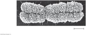
Q2. At what part of the cell cycle would you see a chromosome that looks like this?
Background
Topic: Cell Cycle Phases and Chromosome Structure
This question tests your understanding of when chromosomes are duplicated and condensed during the cell cycle.
Key Terms:
G1 phase: Cell grows and prepares for DNA replication.
S phase: DNA is replicated.
G2 phase: Cell prepares for mitosis.
M phase: Mitosis, where chromosomes are visible and condensed.
Step-by-Step Guidance
Observe the chromosome image and note its X-shaped, condensed appearance.
Recall that chromosomes are duplicated (X-shaped) after DNA replication and before cell division.
Consider which cell cycle phases feature condensed, duplicated chromosomes.
Match the chromosome appearance to the appropriate cell cycle phase from the answer choices.
Try solving on your own before revealing the answer!
Final Answer: G2 phase and M phase
Chromosomes appear X-shaped and condensed after DNA replication (G2) and during mitosis (M phase).
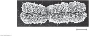
Q3. At what time will the chromosome shown be split into two chromosomes, each the same as the other?
Background
Topic: Mitosis Stages and Chromosome Separation
This question tests your knowledge of the mitotic phases and when sister chromatids are separated.
Key Terms:
Prophase: Chromosomes condense, spindle forms.
Prometaphase: Nuclear envelope breaks down.
Metaphase: Chromosomes align at the cell center.
Anaphase: Sister chromatids separate.
Telophase: Chromosomes reach poles, nuclear envelope reforms.
Step-by-Step Guidance
Review the stages of mitosis and what happens to chromosomes in each stage.
Recall that sister chromatids are joined until a specific stage, then separated.
Identify the stage where the X-shaped chromosome splits into two identical chromosomes.
Match this event to the correct mitotic phase from the answer choices.
Try solving on your own before revealing the answer!
Final Answer: Anaphase
During anaphase, sister chromatids are separated and become individual chromosomes.
Q4. Stained microtubules are marked with a laser during mitosis. How would you explain the results shown?
Background
Topic: Spindle Fiber Dynamics in Mitosis
This question tests your understanding of how spindle fibers move chromosomes during mitosis.
Key Terms:
Microtubules: Protein filaments that form the mitotic spindle.
Centrosome: Organelle that organizes microtubules.
Kinetochore: Protein structure on chromosomes where spindle fibers attach.
Step-by-Step Guidance
Examine the diagram showing microtubule behavior during mitosis.
Recall that microtubules can shorten from either end: the centrosome or the kinetochore.
Analyze the position of the laser mark and how it changes as chromosomes move.
Determine whether microtubules shorten at the centrosome or kinetochore end based on the diagram.
Try solving on your own before revealing the answer!
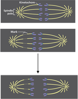
Final Answer: Microtubules shorten at the kinetochore chromosome end.
The laser mark remains stationary, indicating shortening occurs at the kinetochore end.
Q5. For what is cell division used?
Background
Topic: Functions of Cell Division
This question tests your understanding of the biological purposes of cell division in multicellular organisms.
Key Terms:
Growth: Increase in organism size.
Repair: Replacement of damaged or dead cells.
Infection: Not a direct function of cell division.
Step-by-Step Guidance
Recall the main functions of cell division: growth and repair.
Evaluate each answer choice for its relevance to cell division.
Identify which combinations of functions are correct.
Eliminate distractors that do not relate to cell division.
Try solving on your own before revealing the answer!
Final Answer: E. B and C (growth and repair)
Cell division is primarily used for growth and repair in multicellular organisms.
Q6. How does nuclear DNA enter daughter cells?
Background
Topic: DNA Distribution During Cell Division
This question tests your understanding of how genetic material is accurately distributed to daughter cells during mitosis.
Key Terms:
Replication: Copying DNA before cell division.
Chromosome: Structure containing DNA.
Daughter cell: Resulting cell after division.
Step-by-Step Guidance
Recall that DNA is replicated before cell division, resulting in identical copies.
Consider how chromosomes are distributed during mitosis.
Evaluate each answer choice for accuracy regarding DNA distribution.
Eliminate choices that do not match the process of mitosis.
Try solving on your own before revealing the answer!
Final Answer: B. as identical copies of the same chromosomes
Each daughter cell receives identical copies of chromosomes after mitosis.
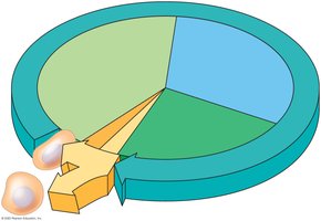
Q7. If you were asked to identify the stage of the cell cycle when the cell completed dividing, which part of the model would you look at?
Background
Topic: Cell Cycle Stages
This question tests your ability to identify the cell cycle phase associated with the completion of cell division.
Key Terms:
Cytokinesis: Division of the cytoplasm, completing cell division.
Mitosis: Division of the nucleus.
S phase: DNA synthesis.
G1 and G2: Growth phases.
Step-by-Step Guidance
Review the cell cycle diagram and identify the phase where two daughter cells are formed.
Recall that cytokinesis follows mitosis and completes cell division.
Match the model's section to the answer choices.
Eliminate phases that do not represent the completion of cell division.
Try solving on your own before revealing the answer!
Final Answer: B. cytokinesis
Cytokinesis is the stage when the cell completes division and two daughter cells are formed.
Q8. If you were asked to identify the stage of the cell cycle when the cell size increases, which part of the model would you look at?
Background
Topic: Cell Growth During the Cell Cycle
This question tests your understanding of when cells grow in size during the cell cycle.
Key Terms:
G1 phase: Cell grows and prepares for DNA replication.
G2 phase: Cell grows and prepares for mitosis.
S phase: DNA synthesis, less growth.
Cytokinesis: Division, not growth.
Step-by-Step Guidance
Review the cell cycle diagram and identify phases associated with cell growth.
Recall that cell size increases during G1 and G2 phases.
Evaluate answer choices for inclusion of these phases.
Eliminate phases not associated with cell growth.
Try solving on your own before revealing the answer!
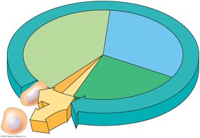
Final Answer: C. G1 and G2 phase
Cell size increases during the G1 and G2 phases of the cell cycle.
Q9. If you saw a cell with condensed chromosomes lined up in the center of the cell, you would know that the cell was in
Background
Topic: Mitosis Stages
This question tests your ability to identify the mitotic phase based on chromosome arrangement.
Key Terms:
Metaphase: Chromosomes align at the cell's equator.
Prophase: Chromosomes condense.
Anaphase: Chromosomes separate.
Step-by-Step Guidance
Recall the sequence of mitosis and the arrangement of chromosomes in each phase.
Identify the phase where chromosomes are lined up at the center (metaphase plate).
Match this description to the answer choices.
Eliminate phases where chromosomes are not aligned at the center.
Try solving on your own before revealing the answer!
Final Answer: C. metaphase
Chromosomes are aligned at the center of the cell during metaphase.
Q10. If you saw a cell with chromosomes starting to condense, you would know that the cell was in
Background
Topic: Mitosis Stages
This question tests your ability to identify the mitotic phase based on chromosome condensation.
Key Terms:
Prophase: Chromosomes begin to condense.
Prometaphase: Chromosomes fully condensed, nuclear envelope breaks down.
Step-by-Step Guidance
Recall the sequence of mitosis and when chromosomes begin to condense.
Identify the phase where condensation starts.
Match this description to the answer choices.
Eliminate phases where chromosomes are already fully condensed or separating.
Try solving on your own before revealing the answer!
Final Answer: A. prophase
Chromosomes begin to condense during prophase.
Q11. If the nuclear envelope has disappeared but the condensed chromosomes are not yet lined up, the cell is in
Background
Topic: Mitosis Stages
This question tests your ability to identify the mitotic phase based on nuclear envelope breakdown and chromosome arrangement.
Key Terms:
Prometaphase: Nuclear envelope disappears, chromosomes not yet aligned.
Metaphase: Chromosomes aligned.
Step-by-Step Guidance
Recall the sequence of mitosis and the events in each phase.
Identify the phase where the nuclear envelope disappears and chromosomes are not yet aligned.
Match this description to the answer choices.
Eliminate phases where chromosomes are aligned or separating.
Try solving on your own before revealing the answer!
Final Answer: B. prometaphase
Prometaphase is when the nuclear envelope disappears and chromosomes are not yet aligned.
Q12. If you saw a cell with chromosomes separating but not yet at the poles, you would know that the cell was in
Background
Topic: Mitosis Stages
This question tests your ability to identify the mitotic phase based on chromosome movement.
Key Terms:
Anaphase: Chromosomes separate and move toward poles.
Telophase: Chromosomes reach poles.
Step-by-Step Guidance
Recall the sequence of mitosis and the movement of chromosomes in each phase.
Identify the phase where chromosomes are separating but not yet at the poles.
Match this description to the answer choices.
Eliminate phases where chromosomes are already at the poles or not yet separated.
Try solving on your own before revealing the answer!
Final Answer: D. anaphase
Chromosomes separate and move toward the poles during anaphase.
Q13. If you wanted to create a drug to stop chromosomes from lining up during mitosis, but otherwise not disturb the overall process, you would want to disrupt/target
Background
Topic: Mitotic Mechanisms and Drug Targets
This question tests your understanding of the structures responsible for chromosome alignment during mitosis.
Key Terms:
Kinetochores: Protein structures on chromosomes where spindle fibers attach.
DNA condensation: Chromosome compaction.
Cytokinesis: Division of cytoplasm.
Step-by-Step Guidance
Recall the process of chromosome alignment during mitosis (metaphase).
Identify the structures involved in attaching chromosomes to spindle fibers.
Evaluate which structure, if disrupted, would prevent alignment but not affect other mitotic processes.
Eliminate choices that would disrupt other aspects of mitosis.
Try solving on your own before revealing the answer!
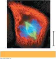
Final Answer: A. kinetochores
Disrupting kinetochores would prevent chromosomes from lining up during mitosis.
Q14. If studying a bacterial cell dividing, you would need to know about
Background
Topic: Prokaryotic Cell Division
This question tests your understanding of how prokaryotes (bacteria) divide, which is different from eukaryotic mitosis.
Key Terms:
Binary fission: Simple cell division in prokaryotes.
Cytokinesis: Division of cytoplasm (in eukaryotes).
Microtubules/microfilaments: Eukaryotic structures, not used in prokaryotes.
Nuclear envelope: Eukaryotic structure, absent in prokaryotes.
Step-by-Step Guidance
Recall that bacteria divide by binary fission, not mitosis.
Identify the process unique to prokaryotes.
Eliminate answer choices related to eukaryotic cell division.
Match the correct process to the answer choices.
Try solving on your own before revealing the answer!
Final Answer: B. binary fission
Bacteria divide by binary fission, not mitosis or cytokinesis.
Q15. A cell has 8 chromosomes during G1. After DNA replication, how many chromosomes and chromatids will the cell contain?
Background
Topic: Chromosome and Chromatid Number After DNA Replication
This question tests your understanding of chromosome and chromatid numbers before and after DNA replication.
Key Terms:
Chromosome: DNA molecule, counted by centromeres.
Chromatid: Each half of a duplicated chromosome.
G1 phase: Before DNA replication.
S phase: DNA replication occurs.
Step-by-Step Guidance
Recall that chromosome number is counted by centromeres, not DNA content.
After DNA replication, each chromosome consists of two sister chromatids.
Calculate the number of chromosomes and chromatids after replication.
Match your calculation to the answer choices.
Try solving on your own before revealing the answer!
Final Answer: 8 chromosomes and 16 chromatids
After DNA replication, the cell has 8 chromosomes (centromeres) and 16 chromatids (DNA molecules).
Q16. A cell contains 10 X-shaped chromosomes during metaphase. How many chromosomes will be present in the entire cell immediately after sister chromatids separate during anaphase?
Background
Topic: Chromosome Number After Chromatid Separation
This question tests your understanding of how chromosome numbers change during mitosis.
Key Terms:
X-shaped chromosome: Duplicated chromosome (two sister chromatids).
Anaphase: Sister chromatids separate.
Step-by-Step Guidance
Recall that each X-shaped chromosome consists of two sister chromatids.
During anaphase, sister chromatids separate and are counted as individual chromosomes.
Calculate the total number of chromosomes after separation.
Match your calculation to the answer choices.
Try solving on your own before revealing the answer!
Final Answer: 20 chromosomes
After sister chromatids separate, each is counted as a chromosome, so there are 20 chromosomes.
Q17. Cell A contains 4 X-shaped chromosomes. Cell B contains 8 I-shaped chromosomes. Which statement is correct?
Background
Topic: Chromosome and DNA Molecule Counting
This question tests your understanding of chromosome and DNA molecule numbers in different cell stages.
Key Terms:
X-shaped chromosome: Duplicated chromosome (two sister chromatids).
I-shaped chromosome: Single chromatid (unduplicated chromosome).
Step-by-Step Guidance
Recall that X-shaped chromosomes have two DNA molecules (sister chromatids).
I-shaped chromosomes have one DNA molecule each.
Compare the number of chromosomes and DNA molecules in each cell.
Evaluate the answer choices for accuracy.
Try solving on your own before revealing the answer!
Final Answer: Cell B has twice as many DNA molecules as Cell A.
Cell A has 4 chromosomes (8 DNA molecules), Cell B has 8 chromosomes (8 DNA molecules).
Q18. A student counts 24 sister chromatids during metaphase. Assuming normal mitosis, how many chromosomes will each daughter cell contain?
Background
Topic: Chromosome Number After Mitosis
This question tests your understanding of chromosome distribution after mitosis.
Key Terms:
Sister chromatid: One of two identical halves of a duplicated chromosome.
Metaphase: Chromosomes aligned, each with two chromatids.
Daughter cell: Resulting cell after division.
Step-by-Step Guidance
Recall that each chromosome consists of two sister chromatids during metaphase.
After mitosis, each daughter cell receives one chromatid from each chromosome.
Calculate the number of chromosomes in each daughter cell.
Match your calculation to the answer choices.
Try solving on your own before revealing the answer!
Final Answer: 12 chromosomes
Each daughter cell will contain 12 chromosomes after mitosis.
