 뒤로
Cell Division: The Cell Cycle and Mitosis – Study Notes for General Biology
스터디 가이드 - 스마트 노트
 자료에 맞춘 맞춤형 노트, 핵심 정의, 예시, 맥락을 확장해 제공합니다.
Cell Division and the Cell Cycle
Overview of Cell Division
Cell division is a fundamental process that enables organisms to reproduce, grow, and maintain their tissues. The continuity of life depends on the reproduction of cells, which occurs through cell division. This process is integral to the cell cycle, the series of events from a cell's formation to its own division.
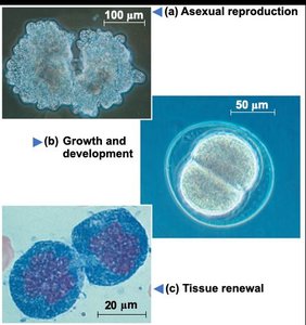
Asexual reproduction: Single-celled organisms reproduce by dividing into two identical cells.
Growth and development: Multicellular organisms grow by increasing their cell number through division.
Tissue renewal: Damaged or old cells are replaced by new cells via division.
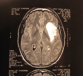
Errors in cell division: Mistakes can lead to diseases such as cancer.
Genetic Material Organization
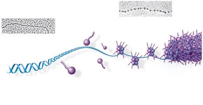
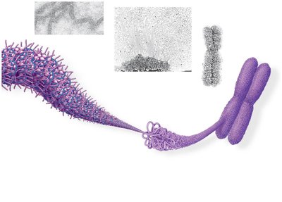
Cells contain genetic material in the form of DNA, which is organized into genes, chromosomes, and chromatin. The packaging of DNA is essential for its function and division.
Gene: Functional unit of DNA containing instructions to make a protein.
Genome: All the DNA in a cell, which may be a single or multiple DNA molecules.
Chromosome: A single DNA molecule, visible when tightly packaged.
Chromatin: Threadlike DNA/protein complex in the nucleus of eukaryotic cells.
Euchromatin: Loosely packed chromatin, easily expressed.
Heterochromatin: Tightly packed chromatin, not easily expressed (e.g., centromeres, telomeres).
Chromosome Number and Structure
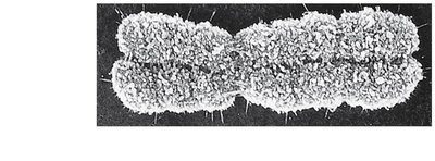
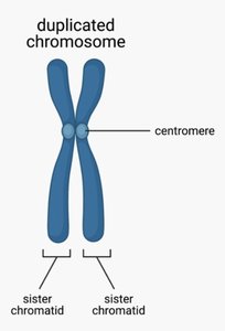
Each species has a characteristic number of chromosomes. Chromosomes duplicate before cell division, forming sister chromatids joined at the centromere.
Diploid number: Total chromosome number in a typical somatic cell.
Sister chromatids: Identical copies of a chromosome, joined at the centromere.
Species | Common Name | Diploid # |
|---|---|---|
Homo sapiens | Human | 46 |
Pan troglodytes | Chimpanzee | 48 |
Canis familiaris | Dog | 78 |
Felis domesticus | Cat | 38 |
Mus musculus | Mouse | 26 |
Rana pipiens | Toad | 26 |
Drosophila melanogaster | Fruitfly | 8 |
Ascaris megalocephala | Roundworm | 2 |
Planaria torva | Flatworm | 16 |
Zea mays | Corn | 20 |
S. cerevisiae | Yeast | 32 |
The Cell Cycle
Phases of the Cell Cycle
The cell cycle consists of interphase and the M phase. Interphase includes G1, S, and G2 phases, while the M phase includes mitosis and cytokinesis.
G1 phase: Cell growth and preparation for DNA replication.
S phase: DNA synthesis and chromosome duplication.
G2 phase: Preparation for mitosis.
M phase: Division of genetic material (mitosis) and cytoplasm (cytokinesis).
Chromosome Duplication and Distribution
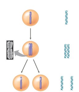
During the S phase, chromosomes are duplicated, resulting in two sister chromatids per chromosome. These chromatids are separated during mitosis, ensuring each daughter cell receives an identical set of chromosomes.
Mitosis: Stages and Mechanisms
Stages of Mitosis (PMAT)
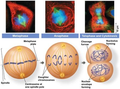
Mitosis is the process by which a cell divides its genetic material. It consists of four main stages: prophase, metaphase, anaphase, and telophase, followed by cytokinesis.
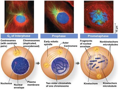
Prophase: Chromosomes condense, sister chromatids are visible, centrosomes separate, mitotic spindle forms, nuclear envelope breaks down.
Metaphase: Chromosomes align on the metaphase plate, spindle fibers attach to kinetochores.
Anaphase: Sister chromatids are pulled apart to opposite poles.
Telophase: Nuclear envelope reforms, chromosomes decondense, cytokinesis begins.
Cytokinesis: Division of cytoplasm, forming two daughter cells.
Cytokinesis in Animal and Plant Cells
Cytokinesis differs between animal and plant cells. Animal cells form a cleavage furrow, while plant cells form a cell plate that develops into a new cell wall.
Regulation of the Cell Cycle
Cell Cycle Checkpoints
The cell cycle is regulated by checkpoints that ensure proper division. These checkpoints occur at G1, G2, and M phases, and require external signals for progression.
G1 checkpoint: Determines if the cell will divide; entry into G0 phase if not.
G2 checkpoint: Ensures chromosomes are duplicated and resources are sufficient.
M checkpoint: Ensures all chromosomes are aligned and attached to the spindle.
External Signals and Cancer
Cell division is influenced by external signals such as anchorage dependence and density-dependent inhibition. Cancer cells often ignore these signals, leading to uncontrolled proliferation.
Anchorage dependence: Cells require a surface to divide.
Density-dependent inhibition: Cells stop dividing when a single layer is formed.
Cancer cells: Divide uncontrollably, forming tumors.
Prokaryotic Cell Division: Binary Fission
Binary Fission in Prokaryotes
Prokaryotes reproduce by binary fission, a simpler process than mitosis. The cell duplicates its DNA and divides into two identical cells.
DNA replication: Circular DNA is copied.
Cell division: Cell splits into two daughter cells.
Summary Table: Cell Turnover Times
Cell Turnover in Different Tissues
Different cell types have varying turnover times, reflecting their roles in growth, maintenance, and repair.
Cell Type | Turnover Time |
|---|---|
Small intestine epithelium | 2-4 days |
Blood Neutrophils | 1-5 days |
Skin epidermis cells | 20-30 days |
Fat cells | 8 years |
Oocytes (female gametes) | Lifetime |
Cardiomyocytes | 0.5% per year |
Central nervous system | Lifetime |
Skeleton | 10% per year |
Key Terms and Concepts
Mitotic spindle: Microtubule structure that separates chromosomes during mitosis.
Centromere: Region joining sister chromatids.
Kinetochore: Protein structure on centromere where spindle fibers attach.
Cleavage furrow: Indentation in animal cells during cytokinesis.
Cell plate: Structure in plant cells that forms the new cell wall during cytokinesis.
Equations and Academic Context
Chromosome number after division:
Example: If there are 20 centromeres at metaphase, each daughter cell will have 10 chromosomes after cytokinesis.
Additional info: The cell cycle is tightly regulated to prevent errors that can lead to diseases such as cancer. Understanding the mechanisms of cell division is fundamental to biology and medicine.
