 뒤로
뒤로Cell Structure, Membrane Function, Metabolism, and Photosynthesis: Study Guide for General Biology
스터디 가이드 - 스마트 노트
 자료에 맞춘 맞춤형 노트, 핵심 정의, 예시, 맥락을 확장해 제공합니다.
Cell Theory and Cell Types
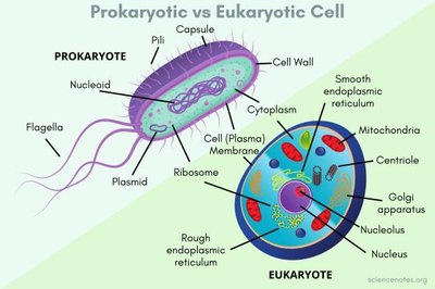
Prokaryotic vs. Eukaryotic Cells
The cell theory states that all living organisms are composed of cells, which are the basic unit of life. Cells are classified as prokaryotic or eukaryotic based on their structural features.
Prokaryotic Cells: Smaller, simpler, lack a nucleus, and do not have membrane-bound organelles. Found in Bacteria and Archaea.
Eukaryotic Cells: Larger, contain a nucleus, and possess membrane-bound organelles. Found in protists, fungi, animals, and plants.
Key Differences: Eukaryotes have compartmentalized functions, while prokaryotes carry out all functions in a single space.
Cell Organelles and Their Functions
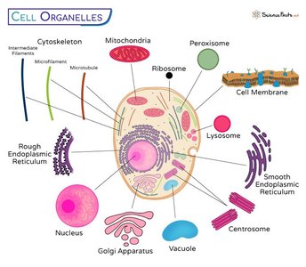
Major Organelles in Eukaryotic Cells
Eukaryotic cells contain specialized organelles that perform distinct functions necessary for cellular life.
Cell Membrane: Phospholipid bilayer that regulates entry and exit of substances.
Vacuole: Stores water, food, and maintains osmotic pressure.
Nucleus: Contains genetic material; site of transcription.
Nuclear Envelope: Double membrane, selectively permeable.
Nucleoplasm: Gelatinous substance protecting genetic material.
Nucleolus: Dense region for ribosome synthesis.
Chromatin: DNA-protein complex involved in cell division.
Lysosome: Digests unwanted substances; absent in plant cells.
Endoplasmic Reticulum (ER): Rough ER synthesizes proteins; Smooth ER synthesizes lipids and carbohydrates.
Ribosomes: Protein factories; composed of rRNA and proteins.
Golgi Apparatus: Modifies, packages, and transports proteins and lipids.
Mitochondria: Powerhouse; site of ATP production and cellular respiration.
Peroxisomes: Detoxify and participate in lipid metabolism.
Cytoskeleton: Network of protein filaments for shape and support.
Centrosome: Organizes microtubules during cell division.
Plant vs. Animal Cells
Structural Differences
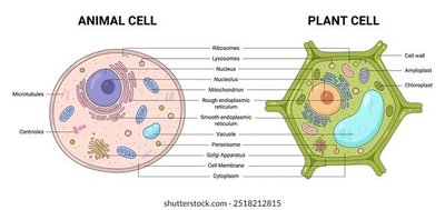
Plant and animal cells share many organelles but differ in certain structures and functions.
Plant Cells: Have a cell wall, chloroplasts, and large central vacuole.
Animal Cells: Lack cell wall and chloroplasts, have smaller vacuoles, and contain centrioles.
Cell Fractionation
Purpose and Process
Cell fractionation separates organelles to study their individual functions, providing insight into cellular operations such as energy production and protein synthesis.
Surface Area and Volume in Cell Size
Importance of Surface Area-to-Volume Ratio
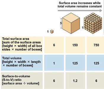
The surface area-to-volume ratio affects the efficiency of material exchange in cells. Smaller cells have higher ratios, facilitating transport and communication.
As cell size increases, volume grows faster than surface area, limiting exchange efficiency.
Cytoskeleton Components
Types and Functions
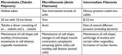
The cytoskeleton is composed of three main types of fibers, each with distinct roles in cell structure and movement.
Type | Structure | Function |
|---|---|---|
Microtubules | Hollow tubes (α- and β-tubulin) | Cell shape, motility, chromosome movement |
Microfilaments | Actin strands | Cell shape, contraction, movement |
Intermediate Filaments | Fibrous proteins | Structural support, anchorage |
Central Dogma and Protein Synthesis
From DNA to Secreted Protein
The central dogma describes the flow of genetic information: DNA → RNA → Protein. Secreted proteins follow a pathway through the endomembrane system.
Transcription: DNA is transcribed to mRNA in the nucleus.
RNA Processing: Pre-mRNA is modified to mature mRNA.
Translation: mRNA is translated into polypeptide on ribosomes.
Secretory Pathway: Proteins are processed in ER and Golgi, then secreted.
Intercellular Junctions
Types and Roles
Cells communicate and adhere via specialized junctions.
Plasmodesmata: Channels in plant cell walls for transport.
Tight Junctions: Prevent leakage between cells.
Desmosomes: Anchor cells together.
Gap Junctions: Allow cytoplasmic exchange.
Membrane Structure and Function
Fluid Mosaic Model
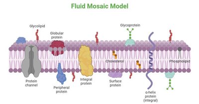
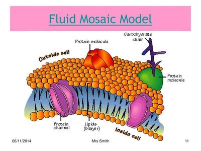
The plasma membrane is a dynamic structure composed of a phospholipid bilayer with embedded proteins, cholesterol, and carbohydrates.
Phospholipids: Form the bilayer, amphipathic.
Proteins: Integral and peripheral, perform transport and signaling.
Cholesterol: Modulates fluidity.
Carbohydrates: Cell recognition.
Membrane Proteins
Functions and Types
Membrane proteins are essential for transport, signaling, enzymatic activity, and structural support.
Integral Proteins: Span the bilayer, amphipathic.
Peripheral Proteins: Attach to surfaces, support and regulate.
Transport Across Membranes
Modes of Transport
Cells use various mechanisms to move substances across membranes.
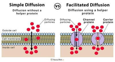
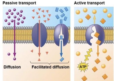
Simple Diffusion: Movement of small, nonpolar molecules without proteins.
Facilitated Diffusion: Uses channel or carrier proteins for larger or charged molecules.
Passive Transport: No energy required; substances move down concentration gradients.
Active Transport: Requires ATP; moves substances against gradients.
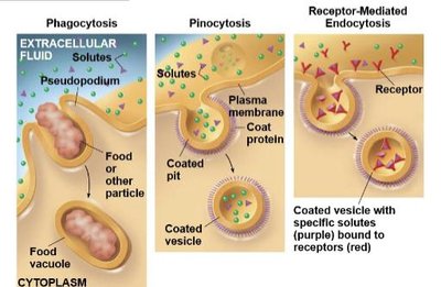
Bulk Transport: Endocytosis and exocytosis move large molecules.
Bulk Transport Types
Phagocytosis: Cellular eating.
Pinocytosis: Cellular drinking.
Receptor-mediated Endocytosis: Specific molecule uptake.
Osmosis and Tonicity
Hypertonic, Hypotonic, Isotonic Solutions
Osmosis is the movement of water across membranes, influenced by solute concentration.
Hypertonic: Water leaves cell; cell shrinks.
Hypotonic: Water enters cell; cell swells.
Isotonic: No net water movement; cell remains stable.
Sodium-Potassium Pump
Mechanism and Importance
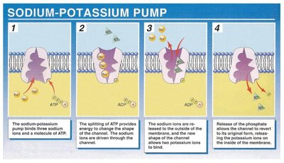
The sodium-potassium pump actively transports Na+ out and K+ into the cell, maintaining ion gradients and membrane potential.
Uses ATP for energy.
Essential for nerve impulses and cell volume regulation.
Metabolism and Energy
Metabolism, Anabolism, Catabolism, Endergonic, Exergonic
Metabolism encompasses all chemical reactions in an organism, divided into anabolic (building) and catabolic (breaking down) pathways.
Endergonic: Requires energy input ().
Exergonic: Releases energy ().
Thermodynamics
First and Second Laws
The first law states energy is conserved; the second law states entropy increases with energy transformations.
Evolutionary Significance of Mitochondria and Chloroplasts
Endosymbiotic Theory
Mitochondria and chloroplasts originated from free-living bacteria, becoming essential for energy production and photosynthesis in eukaryotes.
Energy Flow in Biological Systems
Tracking Energy from the Sun
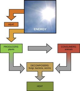
Energy flows from the sun to producers (plants), then to consumers (animals), and finally to decomposers, with heat lost at each step.
ATP and ADP Structure and Function
Energy Transfer Molecules
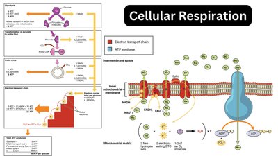
ATP (three phosphates) is the main energy carrier; ADP (two phosphates) is recycled to ATP. ATP powers cellular processes, and is produced in mitochondria and chloroplasts.
Enzymes and Regulation
Enzyme Specificity and Regulation
Enzymes are protein catalysts with specific active sites determined by tertiary structure. Their activity is affected by pH, temperature, cofactors, coenzymes, and inhibitors.
Competitive Inhibitors: Block active site.
Non-competitive Inhibitors: Alter enzyme shape.
Cofactors: Inorganic helpers.
Coenzymes: Organic helpers.
Redox Reactions in Cellular Respiration
Oxidation and Reduction
Redox reactions transfer electrons, powering ATP synthesis. Glucose is oxidized, electron carriers (NADH, FADH2) are reduced, and deliver electrons to the electron transport chain.
Cellular Respiration
Stages and Locations
Cellular respiration occurs in three stages: glycolysis (cytoplasm), Krebs cycle (mitochondrial matrix), and electron transport chain (inner mitochondrial membrane).
Glycolysis: Glucose → pyruvate, produces ATP and NADH.
Krebs Cycle: Pyruvate → CO2, produces ATP, NADH, FADH2.
Electron Transport Chain: NADH/FADH2 → ATP, O2 is final electron acceptor.
ATP Synthase and Chemiosmosis
Role of H+ Gradient
ATP synthase uses the proton gradient across the inner mitochondrial membrane to synthesize ATP during oxidative phosphorylation.
Alternatives to Aerobic Respiration
Anaerobic Respiration and Fermentation
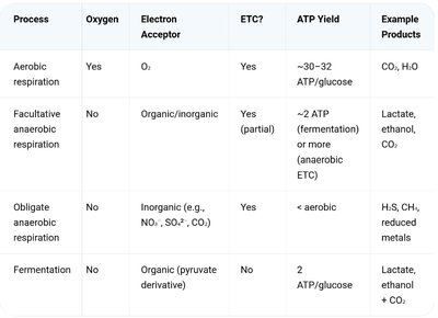
Cells can generate ATP without oxygen via anaerobic respiration or fermentation, using alternative electron acceptors or regenerating NAD+ through pyruvate reduction.
Process | Oxygen | Electron Acceptor | ETC? | ATP Yield | Example Products |
|---|---|---|---|---|---|
Aerobic respiration | Yes | O2 | Yes | ~30–32 ATP/glucose | CO2, H2O |
Facultative anaerobic respiration | No | Organic/inorganic | Yes (partial) | ~2 ATP | Lactate, ethanol, CO2 |
Obligate anaerobic respiration | No | Inorganic | Yes | < aerobic | H2S, CH4, reduced metals |
Fermentation | No | Organic (pyruvate derivative) | No | 2 ATP/glucose | Lactate, ethanol + CO2 |
Chemiosmosis in Mitochondria and Chloroplasts
Differences and Similarities
Chemiosmosis uses electron transport to create a proton gradient, driving ATP synthesis. In mitochondria, electrons from NADH/FADH2 reduce O2; in chloroplasts, electrons from water are energized by light.
Absorbance vs. Action Spectrum
Comparison and Significance
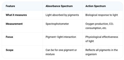
Absorbance spectrum measures light absorption by pigments; action spectrum measures biological response to light. They may differ due to pigment composition and physiological factors.
Feature | Absorbance Spectrum | Action Spectrum |
|---|---|---|
What it measures | Light absorbed by pigments | Biological response to light |
Measurement | Spectrophotometer | Oxygen production, CO2 consumption, etc. |
Focus | Pigment–light interaction | Physiological effectiveness of light |
Scope | Can be for one pigment or mixture | Reflects all pigments in the organism |
Electromagnetic Spectrum and Photosynthesis
Photosynthetically Active Radiation
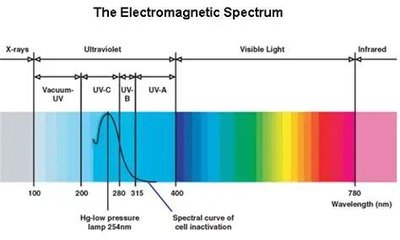
Plants use light in the 400–700 nm range for photosynthesis, corresponding to visible light.
Photosynthesis: Stages and Pathways
Light-Dependent Reactions and Calvin Cycle
Photosynthesis occurs in two stages: light-dependent reactions (thylakoid membranes) produce ATP, NADPH, and O2; Calvin cycle (stroma) uses ATP and NADPH to fix CO2 into glucose.
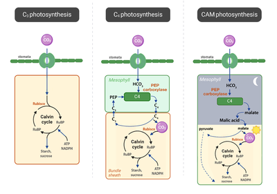
C3, C4, and CAM Pathways
Plants use different pathways to fix CO2 and minimize photorespiration.
C3: Direct fixation by Rubisco; efficient in cool, moist environments.
C4: Spatial separation; PEP carboxylase fixes CO2 in mesophyll, then Calvin cycle in bundle-sheath cells; adapted to hot, dry climates.
CAM: Temporal separation; CO2 fixed at night, used during day; adapted to arid environments.
Relationship Between Photosynthesis and Cellular Respiration
Complementary Processes
Photosynthesis stores energy in glucose; cellular respiration releases energy from glucose. Both are essential for energy flow in living systems.
