 뒤로
Cellular Energetics and Photosynthesis: General Biology Study Guide
스터디 가이드 - 스마트 노트
 자료에 맞춘 맞춤형 노트, 핵심 정의, 예시, 맥락을 확장해 제공합니다.
Cellular Energetics
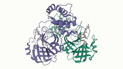
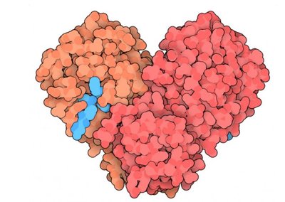
Enzyme Structure
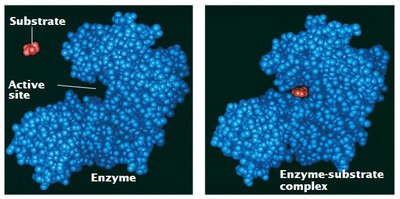
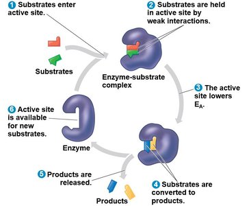
Enzymes are specialized proteins that catalyze biochemical reactions in living organisms. Their three-dimensional structure is essential for their function, particularly the active site, which is the region where substrates bind and reactions occur.
Enzyme: A protein folded into a specific shape with an active site.
Active Site: The location on the enzyme where substrate molecules bind.
Substrate: The reactant that an enzyme acts upon.
Enzyme-Substrate Complex: Formed when the enzyme binds to its substrate.
Substrate Specificity of Enzymes
Enzymes are highly specific, only catalyzing reactions for particular substrates. This specificity is due to the precise shape and chemical environment of the active site.
Substrate: The molecule upon which an enzyme acts.
Enzyme-Substrate Complex: Temporary association between enzyme and substrate.
Enzyme Catalysis
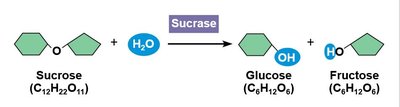
Enzymes act as catalysts, speeding up chemical reactions without being consumed. Most enzyme names end in "-ase" (e.g., sucrase).
Catalyst: A substance that increases the rate of a reaction without being used up.
Example: Sucrase hydrolyzes sucrose into glucose and fructose.
Enzymes Lower Activation Energy
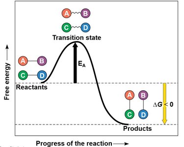
Enzymes lower the activation energy (EA) required for reactions, making them proceed faster. They achieve this by orienting substrates, straining bonds, providing a favorable microenvironment, or forming temporary covalent bonds.
Activation Energy (EA): The energy barrier that must be overcome for a reaction to occur.
Mechanisms: Substrate orientation, bond strain, microenvironment, covalent bonding.
Environmental Impact on Enzyme Function
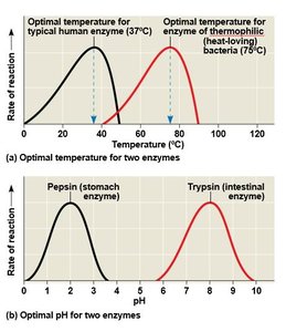
Enzyme activity is influenced by environmental factors such as temperature, pH, and the presence of chemicals. The rate of reaction also depends on enzyme and substrate concentrations, cofactors, coenzymes, and inhibitors.
Temperature: Each enzyme has an optimal temperature.
pH: Each enzyme has an optimal pH range.
Cofactors/Coenzymes: Non-protein helpers required for enzyme activity.
Inhibitors: Molecules that decrease enzyme activity.
Enzyme Inhibitors
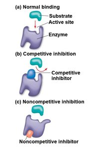
Enzyme inhibitors can be competitive or noncompetitive. Competitive inhibitors bind to the active site, blocking substrate access. Noncompetitive inhibitors bind elsewhere, altering the enzyme's shape and reducing activity.
Competitive Inhibitor: Competes with substrate for active site.
Noncompetitive Inhibitor: Binds to another site, changing enzyme shape.
Allosteric Regulation and Cooperativity
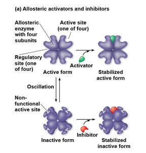
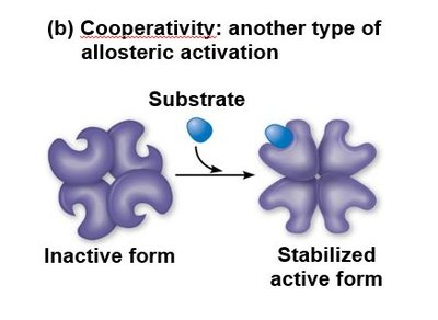
Allosteric enzymes have multiple subunits and can be regulated by activators or inhibitors binding to sites other than the active site. Cooperativity is a form of allosteric regulation where substrate binding to one active site enhances activity at other sites.
Allosteric Activation: Stabilizes active form of enzyme.
Allosteric Inhibition: Stabilizes inactive form.
Cooperativity: Substrate binding amplifies enzyme activity.
Feedback Inhibition
Feedback inhibition occurs when the end product of a metabolic pathway inhibits an earlier step, preventing overproduction and conserving resources.
Feedback Inhibition: End product shuts down pathway.
Cellular Energy
Metabolism and Metabolic Pathways
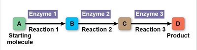
Metabolism is the sum of all chemical reactions in an organism. Metabolic pathways are sequences of reactions, each catalyzed by a specific enzyme, leading from a starting molecule to a product.
Catabolic Pathways: Break down molecules, releasing energy.
Anabolic Pathways: Build molecules, consuming energy.
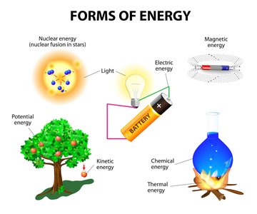
Forms of Energy
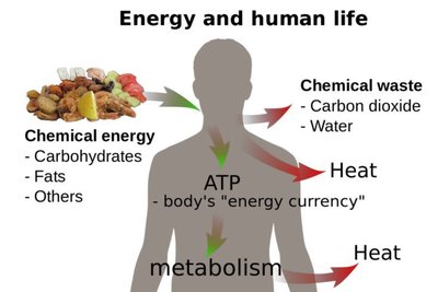
Energy is the capacity to cause change and exists in various forms, including kinetic, potential, chemical, and thermal energy. Energy can be converted from one form to another.
Kinetic Energy: Energy of motion.
Thermal Energy: Kinetic energy of atoms/molecules.
Potential Energy: Energy due to position or structure.
Chemical Energy: Potential energy in chemical bonds.
Thermodynamics
Thermodynamics is the study of energy transformations. The first law states that energy cannot be created or destroyed, only transferred or transformed. The second law states that every energy transfer increases entropy (disorder).
First Law: Conservation of energy.
Second Law: Entropy increases with energy transfer.
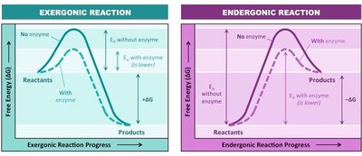
Exergonic and Endergonic Reactions
Exergonic reactions release free energy and are spontaneous. Endergonic reactions absorb free energy and are nonspontaneous.
Exergonic:
Endergonic:
Oxidation-Reduction (Redox) Reactions
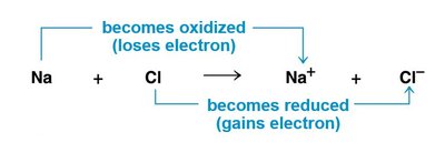
Redox reactions involve the transfer of electrons. Oxidation is the loss of electrons, while reduction is the gain of electrons.
Oxidation: Loses electrons (or gains oxygen/loses hydrogen).
Reduction: Gains electrons (or gains hydrogen).
ATP: The Cell’s Energy Source
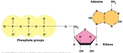
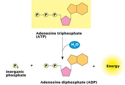
ATP (adenosine triphosphate) is the primary energy carrier in cells. It consists of ribose, adenine, and three phosphate groups. Energy is released when the terminal phosphate bond is broken by hydrolysis.
ATP Structure: Ribose, adenine, three phosphates.
ATP Hydrolysis: Releases energy for cellular work.
Role of ATP in Cell Metabolism
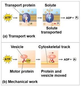
ATP powers three main types of cellular work: chemical, transport, and mechanical. Energy coupling uses exergonic reactions to drive endergonic ones, mediated by ATP hydrolysis.
Chemical Work: Drives endergonic reactions.
Transport Work: Pumps substances across membranes.
Mechanical Work: Moves cellular structures.
Photosynthesis
Overview of Photosynthesis
Photosynthesis converts solar energy into chemical energy. It occurs in plants, algae, and some prokaryotes. Autotrophs sustain themselves without consuming other organisms; photoautotrophs use sunlight to make organic molecules from water and carbon dioxide.
Photosynthesis: Solar energy to chemical energy.
Autotrophs: Self-sustaining organisms.
Photoautotrophs: Use sunlight for photosynthesis.
Photosynthesis in Plants
Photosynthesis primarily occurs in leaves, where chlorophyll absorbs light energy. Stomata are pores that allow CO2 to enter and O2 to exit. Chloroplasts, found in mesophyll cells, contain thylakoids (membranes with chlorophyll) and stroma (fluid surrounding thylakoids).
Chlorophyll: Green pigment absorbing light.
Stomata: Pores for gas exchange.
Thylakoids: Membranes with chlorophyll.
Stroma: Fluid surrounding thylakoids.
Photosynthesis Reaction Overview
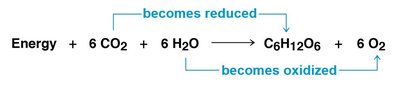
The overall reaction for photosynthesis is:
Light Reactions and Calvin Cycle
Photosynthesis consists of light reactions (in thylakoids) and the Calvin cycle (in stroma). Light reactions split water, release O2, produce ATP, and form NADPH. The Calvin cycle uses ATP and NADPH to fix carbon and produce sugars.
Light Reactions: Split water, release O2, produce ATP and NADPH.
Calvin Cycle: Fixes CO2, forms sugar (G3P).
Role of Pigments in Photosynthesis
Pigments absorb visible light. Chlorophyll a is the main pigment; accessory pigments like chlorophyll b and carotenoids broaden the spectrum and protect chlorophyll from excess light.
Chlorophyll a: Main photosynthetic pigment.
Accessory Pigments: Broaden absorption spectrum.
Photosystems and Electron Flow
Photosystems are complexes of pigments and proteins that funnel light energy to a reaction center. There are two types: Photosystem II (absorbs 680 nm) and Photosystem I (absorbs 700 nm). Electron flow can be cyclic or noncyclic, producing ATP and NADPH.
Photosystem II: Functions first, absorbs 680 nm.
Photosystem I: Absorbs 700 nm.
Noncyclic Electron Flow: Produces ATP and NADPH.
Cyclic Electron Flow: Produces only ATP.
Calvin Cycle Details
The Calvin cycle regenerates its starting material and builds sugar from CO2 using ATP and NADPH. It has three phases: carbon fixation (catalyzed by rubisco), reduction, and regeneration of the CO2 acceptor (RuBP).
Carbon Fixation: Incorporates CO2 into organic molecules.
Reduction: Uses ATP and NADPH.
Regeneration: Restores RuBP for next cycle.
Problems Plants Face: Photorespiration
Plants may close stomata to conserve water, limiting photosynthesis and causing O2 buildup. Photorespiration occurs when rubisco adds O2 instead of CO2, consuming energy and reducing efficiency.
Photorespiration: Rubisco adds O2, reducing photosynthetic output.
Plant Types: C3, C4, and CAM Plants
C3 plants fix CO2 into a three-carbon compound but are susceptible to photorespiration. C4 plants minimize photorespiration by incorporating CO2 into four-carbon compounds. CAM plants open stomata at night, storing CO2 as organic acids for use during the day.
C3 Plants: Most common, susceptible to photorespiration.
C4 Plants: Minimize photorespiration, use four-carbon compounds.
CAM Plants: Open stomata at night, store CO2 as acids.
Cellular Respiration
Overview of Cellular Respiration
Cellular respiration is the process by which cells extract energy from glucose. It involves four stages: glycolysis, pyruvate oxidation, citric acid cycle, and oxidative phosphorylation.
Glycolysis: Breaks down glucose into pyruvate.
Pyruvate Oxidation: Converts pyruvate to acetyl CoA.
Citric Acid Cycle: Completes glucose breakdown.
Oxidative Phosphorylation: Synthesizes most ATP.
Glycolysis
Glycolysis occurs in the cytoplasm and consists of two phases: energy investment and energy payoff. It produces two molecules of pyruvate from one glucose.
Energy Investment Phase: Uses ATP to phosphorylate glucose.
Energy Payoff Phase: Produces ATP and NADH.
Pyruvate Oxidation
In the presence of O2, pyruvate enters the mitochondrion and is converted to acetyl CoA through three reactions: oxidation, reduction of NAD+, and combination with coenzyme A.
Oxidation: Releases CO2.
Reduction: NAD+ to NADH.
Acetyl CoA Formation: Combines two-carbon fragment with coenzyme A.
Citric Acid Cycle
The citric acid cycle has eight steps, starting with acetyl CoA joining oxaloacetate to form citrate. The cycle produces NADH and FADH2, which relay electrons to the electron transport chain.
Electron Carriers: NADH and FADH2.
ATP Synthesis: Powered by oxidative phosphorylation.
Electron Transport Chain and Chemiosmosis
The electron transport chain is located in the cristae of the mitochondrion. Electrons are passed through protein complexes, ultimately reducing O2 to water. Chemiosmosis uses the H+ gradient to drive ATP synthesis via ATP synthase.
Electron Transport Chain: Transfers electrons, releases energy.
Chemiosmosis: H+ gradient powers ATP synthesis.
Fermentation
Fermentation allows cells to generate ATP without oxygen. Alcohol fermentation converts pyruvate to ethanol, releasing CO2. Lactic acid fermentation reduces pyruvate to lactate, used by fungi, bacteria, and human muscle cells.
Alcohol Fermentation: Pyruvate to ethanol, CO2 released.
Lactic Acid Fermentation: Pyruvate to lactate, no CO2 released.
Fitness and Variation
Variation and Fitness
Variation refers to differences in type, structure, or molecules among living things. Increased variation enhances fitness, defined as an organism's ability to survive and reproduce in its environment.
Fitness: Ability to survive and reproduce.
Variation: Differences in cells, molecules, etc.
Variation in Phospholipids
Cell membranes contain various lipid types, such as cholesterol (maintains fluidity) and phosphatidylcholine (most abundant phospholipid).
Variation in Hemoglobin
Hemoglobin carries oxygen in blood. Fetal hemoglobin (Hemoglobin F) binds oxygen strongly for efficient transfer in the placenta, while adult hemoglobin (Hemoglobin A) releases oxygen more easily to cells.
Variation in Chlorophyll/Pigments
Chlorophyll varies among photosynthetic organisms, with different forms (A, B, C, etc.) absorbing different wavelengths of light, enhancing photosynthetic efficiency.
Chlorophyll A: Found in all plants.
Chlorophyll B-E: Found in various groups.
