 뒤로
Chapter 4: The Cell – Structure, Function, and Microscopy
스터디 가이드 - 스마트 노트
 자료에 맞춘 맞춤형 노트, 핵심 정의, 예시, 맥락을 확장해 제공합니다.
The Cell: Historical Foundations and Cell Theory
Historical Figures in Cell Biology
The study of cells began with the invention and use of microscopes, leading to foundational discoveries in biology.
Robert Hooke (1665): Used a microscope to view cork and coined the term "cell."
Anton van Leeuwenhoek (1674): Improved lenses and observed living cells and microorganisms.
Matthias Schleiden (1838): Concluded all plants are made of cells.
Theodor Schwann (1839): Concluded all animals are made of cells.
Rudolph Virchow (1855): Stated all cells arise from pre-existing cells.
The Cell Theory
Cell theory is a fundamental concept in biology, stating:
All living things are composed of one or more cells.
Cells are the basic units of structure and function in organisms.
Cells are produced only from pre-existing cells.
Microscopy: Tools for Studying Cells
Magnification and Resolution
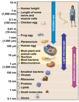
Microscopes are essential for visualizing cells and their structures. Magnification is the ratio of an object's image size to its real size, while resolution is the clarity of the image, defined as the minimum distance two points can be distinguished as separate.
Types of Microscopes
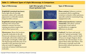
Light Microscopes (LM): Use light to view specimens, magnify up to ~1000X, and can observe living cells.
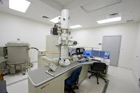
Electron Microscopes (EM): Use electron beams, magnify up to ~1,000,000X, and reveal internal structures but only for dead cells.
TEM (Transmission Electron Microscope): Views internal cell structures.
SEM (Scanning Electron Microscope): Produces 3D images of cell surfaces.
Pros and Cons of Microscopes
EM Advantages: High magnification and resolution; reveals organelles.
EM Disadvantages: Only for dead cells; expensive and large.
LM Advantages: Can study live cells; affordable and portable.
LM Disadvantages: Lower magnification and resolution.
Parts of the Microscope
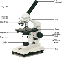
Eyepiece, pointer, body tube, revolving nose piece, stage, stage clips, diaphragm, lamp, arm, fine/coarse focus, base, objectives (scanning, low, high, oil immersion).
Microscope Use and Care
Carry with two hands; keep away from desk edge; clean with lens paper.
Start with lowest power objective; use coarse then fine focus; adjust diaphragm.
Never use coarse focus on high power; avoid 100X objective unless trained.
Cell Structure and Function
Limits Governing Cell Size
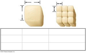
Cell size is limited by transport efficiency. As cells grow, volume increases faster than surface area, reducing efficiency in nutrient and waste exchange.
Small cells have a higher surface area-to-volume ratio, making them more efficient.
Larger organisms have more cells, not larger cells.
Plasma (Cell) Membrane
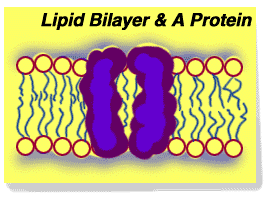
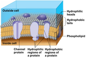
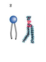
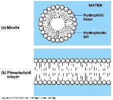
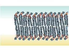
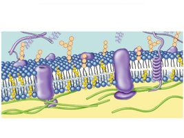
The plasma membrane is a phospholipid bilayer with embedded proteins, forming a selective barrier for the cell.
Encloses cell contents.
Regulates entry and exit of substances.
Fluid-mosaic model: dynamic structure with proteins and cholesterol.
Cell Characteristics
All cells have a plasma membrane, cytosol, DNA (chromosomes), and ribosomes.
Cytoplasm includes cytosol and organelles; contains nutrients and enzymes.
Cell Types: Prokaryote vs. Eukaryote
Cells are classified as prokaryotic or eukaryotic based on internal structure.
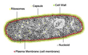
Prokaryotic Cells: No nuclear membrane, no membrane-bound organelles, small (1-10 µm), found in bacteria and archaea.
Eukaryotic Cells: Membrane-bound nucleus and organelles, larger (2-1000 µm), found in protists, fungi, plants, and animals.
Prokaryotic Cell Structure
Nucleoid region (DNA), ribosomes, plasma membrane, cell wall, capsule, pili, flagella.
Ribosomes differ from eukaryotes and are targets for antibiotics.
Eukaryotic Cell Structure
Eukaryotic cells are more complex, with compartmentalized organelles for specialized functions.
Genetic control: Nucleus, ribosomes
Manufacturing, distribution, breakdown: ER, Golgi, lysosomes, vacuoles, peroxisomes
Energy processing: Mitochondria, chloroplasts
Structural support, movement, communication: Cytoskeleton, plasma membrane, cell wall
Plant vs. Animal Cells
Plant Cells: Cell wall, chloroplasts, large central vacuole, plasmodesmata, no lysosomes or centrioles.
Animal Cells: No cell wall or chloroplasts, small vacuoles, lysosomes, centrioles.
Eukaryotic Organelles and Endomembrane System
Nucleus
The nucleus is the control center, containing DNA (chromatin) and enclosed by a nuclear envelope. The nucleolus synthesizes ribosome subunits.
Endoplasmic Reticulum (ER)
Smooth ER: No ribosomes; synthesizes lipids, breaks down glycogen, processes toxins.
Rough ER: Covered with ribosomes; synthesizes proteins for export or membrane insertion.
Ribosomes
Site of protein synthesis; subunits made in nucleolus.
Free ribosomes synthesize cytosolic proteins; bound ribosomes synthesize membrane/exported proteins.
Golgi Apparatus
Stacks of flattened sacs that modify, sort, and package ER products into vesicles for export or membrane incorporation.
Lysosomes
Membranous sacs containing digestive enzymes; fuse with food vacuoles or damaged organelles for intracellular digestion and recycling.
Defects in lysosomal enzymes cause diseases (e.g., Pompe’s, Tay-Sachs).
Vacuoles
Food vacuoles: Store nutrients.
Contractile vacuoles: Expel excess water.
Central vacuole (plants): Storage and water balance.
Peroxisomes
Metabolic compartments with enzymes to break down fatty acids and detoxify compounds; generate and degrade hydrogen peroxide ().
Energy-Converting Organelles
Mitochondria
Site of cellular respiration; double membrane with highly folded inner membrane (cristae); produces ATP from sugars.
Chloroplasts
Site of photosynthesis; contains chlorophyll; compartments include stroma, thylakoids, and grana.
Endosymbiosis Theory
Mitochondria and chloroplasts evolved from prokaryotes living within larger cells, evidenced by their double membranes, own DNA, ribosomes, and independent reproduction.
Cell Walls and Surface Structures
Plant Cell Walls
Rigid, cellulose-based walls provide support and protection; plasmodesmata allow communication and transport between cells.
Cytoskeleton and Cell Surfaces
Cytoskeleton
Network of protein fibers organizing cell structure and movement.
Microtubules: Hollow, thickest; shape cell, tracks for motor proteins, form centrioles, cilia, flagella.
Intermediate filaments: Reinforce cell shape, anchor organelles.
Microfilaments: Solid rods, thinnest; support shape, involved in motility.
Cilia and Flagella
Structures for movement; cilia are short and numerous, flagella are longer and fewer. Both are composed of microtubules.
Centrioles
Cylinders of microtubules in animal cells, involved in cell division.
Extracellular Matrix (ECM)
Animal cells have an ECM composed mainly of glycoproteins (especially collagen), providing support, movement, and regulation. ECM connects to the cytoskeleton via integrins.
Cell Junctions in Animal Tissues
Tight junctions: Prevent leakage of extracellular fluid.
Anchoring junctions: Fasten cells together into sheets.
Gap junctions: Channels for communication between cells.
Summary of Key Concepts
Microscopes are essential for understanding cell structure and function.
Cell theory defines the cell as the basic unit of life.
Cell size is limited by surface area-to-volume ratio.
Cell membranes are dynamic, selective barriers.
Prokaryotic and eukaryotic cells differ in complexity and compartmentalization.
Organelles perform specialized functions in eukaryotic cells.
Plant and animal cells have distinct structural features.
Mitochondria and chloroplasts likely evolved by endosymbiosis.
Cytoskeleton and ECM provide internal and external support.
Cell junctions enable communication and structural integrity in tissues.
