 뒤로
Circulatory and Respiratory Systems: Structure, Function, and Disease
스터디 가이드 - 스마트 노트
 자료에 맞춘 맞춤형 노트, 핵심 정의, 예시, 맥락을 확장해 제공합니다.
Circulatory Systems: Features and Functions
Diffusion and Gastrovascular Cavities
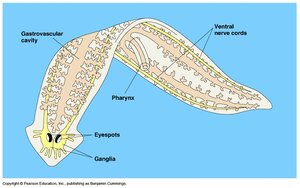
Diffusion is only efficient over small distances, which limits the size and complexity of organisms relying solely on this process. Simple animals, such as flatworms, possess a gastrovascular cavity that is two cell layers thick, allowing direct exchange of substances between cells and the environment.
Gastrovascular cavity: Serves both digestive and circulatory functions in simple animals.
Diffusion: Movement of molecules from high to low concentration; efficient only over microscopic distances.
Example: Flatworms use their gastrovascular cavity for nutrient and gas exchange.
Open vs. Closed Circulatory Systems
Animals have evolved two main types of circulatory systems: open and closed. In open systems, the circulatory fluid (hemolymph) bathes organs directly, while in closed systems, blood is confined to vessels and is distinct from interstitial fluid.
Open circulatory system: Found in arthropods and some mollusks; hemolymph is pumped by the heart into body cavities.
Closed circulatory system: Found in vertebrates and some invertebrates; blood circulates within vessels.
Advantages: Closed systems allow higher pressure and more efficient transport; open systems are energetically cheaper.
Organization of Vertebrate Circulatory Systems
Cardiovascular System Structure
The vertebrate cardiovascular system is a closed system consisting of the heart and blood vessels. Blood flows in one direction through arteries, capillaries, and veins.
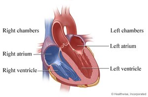
Heart: Composed of atria (collection chambers) and ventricles (pumping chambers).
Arteries: Carry blood away from the heart.
Capillaries: Sites of exchange between blood and interstitial fluid.
Veins: Return blood to the heart.
Blood Flow and Heart Chambers
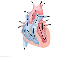
Blood flows through the heart in a specific sequence, passing through the right and left atria and ventricles. The heart's structure ensures efficient circulation and separation of oxygenated and deoxygenated blood.
Right atrium and ventricle: Receive and pump deoxygenated blood to the lungs.
Left atrium and ventricle: Receive and pump oxygenated blood to the body.
The Cardiac Cycle
The cardiac cycle consists of alternating contraction (systole) and relaxation (diastole) phases. Cardiac output is the volume of blood pumped per minute, determined by heart rate and stroke volume.
Systole: Contraction phase; blood is pumped out of the heart.
Diastole: Relaxation phase; heart fills with blood.
Cardiac output:
Heart rate: Number of beats per minute (60-100 bpm).
Stroke volume: Amount of blood pumped per contraction (~70 mL).
Heart Valves and Sounds
Four valves prevent backflow of blood in the heart. The atrioventricular (AV) valves separate atria and ventricles, while semilunar valves control blood flow to the aorta and pulmonary artery. The "lub-dup" sound is caused by the closing of these valves.
AV valves: Prevent backflow into atria.
Semilunar valves: Prevent backflow into ventricles.
Heart murmur: Caused by defective valves.
Maintaining the Heart’s Rhythmic Beat
Some cardiac muscle cells are autorhythmic. The sinoatrial (SA) node acts as the pacemaker, setting the rate and timing of contractions. Impulses travel to the atrioventricular (AV) node and Purkinje fibers, coordinating the heartbeat.
SA node: Initiates electrical impulses.
AV node: Delays impulses before sending them to ventricles.
Purkinje fibers: Distribute impulses to ventricles.
Regulation: Controlled by sympathetic and parasympathetic nervous systems.
Blood: Components and Functions
Blood Composition
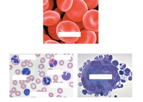
Blood is a connective tissue consisting of cells suspended in plasma. Cellular elements include erythrocytes (red blood cells), leukocytes (white blood cells), and platelets.
Plasma: Liquid matrix containing water, proteins, nutrients, and waste.
Cellular components: Make up ~45% of blood volume.
Erythrocytes (Red Blood Cells)
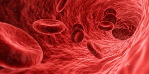
Erythrocytes are the most numerous blood cells and contain hemoglobin, which binds and transports oxygen. Mature erythrocytes lack nuclei and mitochondria.
Hemoglobin: Iron-containing protein; each molecule binds up to 4 O2 molecules.
Function: Oxygen transport.
Leukocytes (White Blood Cells)
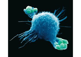
Leukocytes are involved in defense and immunity. There are five major types: monocytes, neutrophils, basophils, eosinophils, and lymphocytes.
Defense mechanisms: Phagocytosis of bacteria and debris; immune responses against foreign substances.
Stem Cells and Blood Cell Replacement
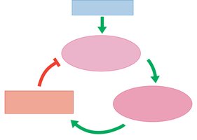
All blood cells develop from stem cells in the red bone marrow. The hormone erythropoietin (EPO) stimulates erythrocyte production when oxygen delivery is low.
Stem cells: Located in ribs, vertebrae, sternum, and pelvis.
EPO: Used clinically to treat anemia.
Blood Clotting
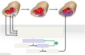
Platelets are cell fragments that function in blood clotting. Coagulation involves a cascade of reactions converting fibrinogen to fibrin, forming a clot. A thrombus is a clot formed within a blood vessel.
Platelets: Initiate clotting.
Fibrin: Forms the meshwork of a clot.
Clotting factors: Include calcium and vitamin K.
Cardiovascular Diseases
Atherosclerosis, Heart Attacks, and Stroke
Cardiovascular diseases are disorders of the heart and blood vessels. Atherosclerosis is the hardening of arteries due to plaque buildup, which can lead to heart attacks or strokes.
Atherosclerosis: Inflammation and lipid accumulation in artery walls.
Heart attack: Blockage of coronary arteries causing cardiac muscle death.
Stroke: Blockage or rupture of arteries in the brain.
Angina pectoris: Chest pain from partial blockage.
Risk Factors and Treatment
Cholesterol levels and inflammation are key risk factors. Low-density lipoprotein (LDL) delivers cholesterol to cells, while high-density lipoprotein (HDL) removes excess cholesterol. A high LDL/HDL ratio increases risk.
LDL: "Bad" cholesterol.
HDL: "Good" cholesterol.
Inflammation: Aspirin can reduce risk.
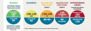
Hypertension (High Blood Pressure)
Hypertension contributes to heart attack, stroke, and other health problems. It can be managed with diet, exercise, and medication.
Normal blood pressure: Less than 120/80 mmHg.
Elevated: 120-129/<80 mmHg.
High blood pressure: 130-139/80-89 mmHg or higher.
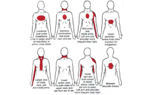
Heart Attack Symptoms in Females
Heart attack symptoms can vary, especially in females. Common symptoms include chest pain, pain in the neck, jaw, or arms, and shortness of breath.
Symptoms: May include pain in unusual locations, fatigue, and nausea.
Gas Exchange and Respiratory Systems
Gas Exchange Mechanisms
Gas exchange is the uptake of oxygen and release of carbon dioxide, occurring by diffusion across specialized respiratory surfaces. These surfaces are large and thin to maximize exchange.
Respiratory surfaces: Skin, gills, tracheae, lungs.
Diffusion: Requires moist, thin membranes.
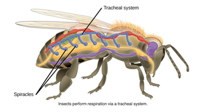
Tracheal Systems in Insects
Insects use a tracheal system, a network of air tubes that open to the outside and reach every cell. Diffusion occurs across moist epithelium, and body movements increase airflow.
Tracheae: Main air tubes.
Spiracles: Openings to the outside.
Lungs and Respiratory Surface Area
Lungs are internal organs with a large surface area for gas exchange. The cells lining respiratory surfaces are thin and moist, facilitating diffusion.
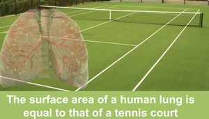
Surface area: Human lungs have a surface area comparable to a tennis court.
Moist membranes: Essential for gas diffusion.
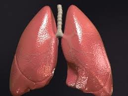
Mammalian Respiratory System Structure
Air enters through the nostrils, is filtered, warmed, and humidified, then passes through the pharynx, larynx, trachea, bronchi, and bronchioles to the alveoli. The epiglottis prevents food from entering the trachea during swallowing.
Pharynx: Directs air and food.
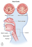
Larynx: Contains vocal cords.
Trachea: Windpipe leading to lungs.
Bronchi and bronchioles: Branching airways.
Vocal Cords and Mucus Escalator
Exhaled air passes over the vocal cords in the larynx to create sounds. Cilia and mucus line the air ducts, moving particles up to the pharynx for removal.
Vocal cords: Produce sound.
Mucus escalator: Cleans respiratory system.
Alveoli and Gas Exchange
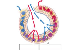
Gas exchange occurs in alveoli, air sacs at the tips of bronchioles. Oxygen diffuses into capillaries, while carbon dioxide diffuses out. Alveoli lack cilia and are coated with surfactant to reduce surface tension.
Alveoli: Main site of gas exchange.
Surfactant: Prevents alveolar collapse.
Preterm babies: May lack surfactant, requiring artificial treatment.
Summary Review
Describe blood: Plasma and cellular components.
Outline blood clotting: Platelet activation, fibrin formation.
Oxygen regulation: EPO and negative feedback.
Cardiovascular diseases: Atherosclerosis, heart attack, stroke, hypertension.
Gas exchange: Respiratory surfaces, tracheal systems, lungs, alveoli.
Additional info: Academic context was added to clarify mechanisms, definitions, and clinical relevance for exam preparation.
