 뒤로
뒤로General Biology Midterm Study Guide: Scientific Method, Chemistry, Biomolecules, Cells, Membranes, Respiration, and Photosynthesis
스터디 가이드 - 스마트 노트
 자료에 맞춘 맞춤형 노트, 핵심 정의, 예시, 맥락을 확장해 제공합니다.
Scientific Method, Experimental Design, and Lab Skills
Overview of the Scientific Method
The scientific method is a systematic approach used to investigate natural phenomena, ensuring reliability and reproducibility of results. It involves several key steps:
Observation and Question: Noticing phenomena and formulating questions.
Hypothesis: A testable explanation that predicts outcomes.
Experiment: Designing and conducting tests to gather data.
Data Analysis: Interpreting results to draw conclusions.
Conclusion: Summarizing findings and their implications.
Peer Review and Publication: Sharing results for validation by the scientific community.
Prediction is the expected outcome, while a hypothesis explains what and why. A theory is a broad, well-supported explanation (e.g., cell theory).
Variables: Independent (changed), dependent (measured), controlled (kept constant).
Controls: Negative (no effect expected), positive (known effect expected).
Lab Safety: Know emergency equipment, report accidents, use proper measurement tools.
Measurement: Use correct units and conversions; density = mass ÷ volume.
Significant Figures: Rules for counting digits in measurements.
Basic Chemistry and Water
Atoms and Elements
Life is composed mainly of a few elements (CHNOPS). Atoms consist of protons, neutrons, and electrons:
Proton: +1 charge, 1 amu, nucleus
Neutron: 0 charge, 1 amu, nucleus
Electron: -1 charge, ~0 amu, orbitals
Atomic number = protons; mass number = protons + neutrons. Isotopes have different neutron counts and may be radioactive (used in dating and medicine).
Chemical Bonds
Nonpolar Covalent: Electrons shared equally (e.g., H2).
Polar Covalent: Unequal sharing, partial charges (e.g., O–H in water).
Ionic: Electron transferred, forming ions (e.g., Na+ Cl-).
Hydrogen Bond: Weak attraction between molecules (e.g., water, DNA).
Van der Waals: Temporary weak attractions.
Electronegativity determines bond polarity. Cations are positive ions; anions are negative.
Water's Properties
Water's polarity and hydrogen bonding give rise to several life-supporting properties:
Cohesion/Adhesion: Water sticks to itself and other surfaces.
High Specific Heat: Stabilizes temperature.
High Heat of Vaporization: Cooling effect (sweating).
Ice Floats: Lower density due to H-bonded lattice.
Universal Solvent: Dissolves polar and ionic substances.
Acids increase [H+], lowering pH; bases decrease [H+] or add OH-, raising pH. Buffers resist pH changes (e.g., blood bicarbonate buffer).
Carbon and Functional Groups
Organic molecules contain carbon. Functional groups determine chemical properties:
Hydroxyl (–OH): Polar, in sugars/alcohols
Carbonyl (C=O): In sugars
Carboxyl (–COOH): Acidic, in amino/fatty acids
Amino (–NH2): Basic, in amino acids
Phosphate (–PO4): In ATP, DNA
Sulfhydryl (–SH): Forms disulfide bridges
Methyl (–CH3): Nonpolar
Biomolecules and Food Macromolecules
Overview of Biomolecules
Three classes are polymers (carbohydrates, proteins, nucleic acids); lipids are not. Dehydration synthesis builds polymers; hydrolysis breaks them down.
Class | Monomer | Bond | Main Roles | Examples |
|---|---|---|---|---|
Carbohydrates | Monosaccharide | Glycosidic | Energy, structure | Glucose, starch, cellulose |
Proteins | Amino acid | Peptide | Enzymes, structure | Albumin, hemoglobin |
Nucleic acids | Nucleotide | Phosphodiester | Genetic info | DNA, RNA |
Lipids | None | Ester | Energy, membranes | Triglycerides, phospholipids |
Carbohydrates
Monosaccharides (e.g., glucose), disaccharides (e.g., maltose), polysaccharides (e.g., starch, cellulose).
Energy storage: starch (plants), glycogen (animals).
Structure: cellulose (plants), chitin (fungi, insects).
Lipids
Hydrophobic; some are amphipathic.
Saturated (solid, straight chains), unsaturated (liquid, kinked chains), trans fats (artificial).
Triglycerides: glycerol + 3 fatty acids.
Phospholipids: hydrophilic head, hydrophobic tails; form cell membranes.
Steroids: four fused rings (e.g., cholesterol).
Proteins
Amino acids (20 types), peptide bonds.
Levels: primary (sequence), secondary (α-helix, β-sheet), tertiary (3D shape), quaternary (multiple polypeptides).
Denaturation: loss of structure/function due to pH, temperature, salt.
Nucleic Acids
Nucleotide = phosphate + pentose sugar + nitrogenous base.
DNA: deoxyribose, bases A/T/C/G, double helix.
RNA: ribose, bases A/U/C/G, single-stranded.
Lab Tests for Macromolecules
Test | Detects | Negative | Positive |
|---|---|---|---|
Benedict's | Reducing sugars | Blue | Green/yellow/orange/red |
Iodine | Starch | Yellow-brown | Blue-black |
Biuret | Protein | Blue | Violet/purple |
Brown-paper | Lipids | Spot dries | Grease spot stays |
Cells, Microscopy, and Subcellular Structures
Microscope Types and Usage
Light microscope: visible light, basic cell structures.
Electron microscope: higher resolution (SEM for surfaces, TEM for internal structures).
Compound microscope: total magnification = ocular × objective.
Proper handling and focusing techniques are essential.
Prokaryotic vs. Eukaryotic Cells
Feature | Prokaryotes | Eukaryotes |
|---|---|---|
Nucleus | None | Membrane-bound |
DNA shape | Circular | Linear |
Organelles | No membrane-bound | Many membrane-bound |
Ribosomes | 70S | 80S |
Size | ~1–10 µm | ~10–100 µm |
Division | Binary fission | Mitosis + cytokinesis |
All cells share a plasma membrane, cytoplasm, ribosomes, and DNA.
Key Organelles and Their Functions
Nucleus: DNA storage, nuclear envelope.
Nucleolus: Ribosome assembly.
Ribosome: Protein synthesis.
Rough ER: Protein folding/modification.
Smooth ER: Lipid synthesis, detoxification.
Golgi apparatus: Modifies, sorts, ships proteins.
Lysosome: Digestion/recycling (mainly animal cells).
Peroxisome: Breaks down H2O2, fatty acids.
Central vacuole: Storage, turgor (plants).
Mitochondrion: Cellular respiration, ATP production.
Chloroplast: Photosynthesis (plants, algae).
Cell wall: Structural support.
Centrioles: Organize microtubules (animal cells).
Endosymbiotic Theory
Mitochondria and chloroplasts originated from engulfed bacteria. Evidence: circular DNA, 70S ribosomes, double membranes, binary fission.
Cytoskeleton and Cell Junctions
Microfilaments: Actin, cell shape, movement.
Intermediate filaments: Keratin, tension, anchoring.
Microtubules: Tubulin, tracks, cilia/flagella, spindle.
Cell junctions: Tight (seal), desmosomes (anchor), gap (communication), plasmodesmata (plants).
Membranes, Energy, Respiration, and Signaling
Membrane Structure and Transport
The fluid mosaic model describes the cell membrane as a phospholipid bilayer with embedded proteins, cholesterol, and carbohydrates. It is selectively permeable.
Simple diffusion: Passive, high to low concentration (O2, CO2).
Facilitated diffusion: Passive, via channels/carriers (glucose).
Osmosis: Water movement toward higher solute concentration.
Active transport: Requires ATP, low to high concentration (Na+/K+ pump).
Endocytosis/Exocytosis: Bulk transport via vesicles.
Tonicity: Hypotonic (water enters), hypertonic (water leaves), isotonic (no net movement).
Energy, Metabolism, and Enzymes
Anabolic: Build molecules, use energy.
Catabolic: Break down molecules, release energy.
Kinetic energy: Motion; potential energy: Stored.
Exergonic: Release energy (); endergonic: Require energy ().
ATP: Adenine + ribose + 3 phosphates; hydrolysis releases energy ().
Enzymes: Protein catalysts, lower activation energy, sensitive to pH/temperature.
Inhibition: Competitive (active site), noncompetitive (allosteric), feedback inhibition.
Cellular Respiration
Cellular respiration converts glucose and oxygen into carbon dioxide, water, and ATP:
Stage | Location | Key Outputs |
|---|---|---|
Glycolysis | Cytoplasm | 2 pyruvate, 2 ATP, 2 NADH |
Pyruvate oxidation | Mitochondrial matrix | Acetyl CoA, CO2, NADH |
Citric acid cycle | Mitochondrial matrix | CO2, NADH, FADH2, 2 ATP |
Electron transport chain | Inner mitochondrial membrane | ~26–34 ATP, H2O |
Without O2, fermentation regenerates NAD+ (lactic acid or alcohol + CO2).
Cell Communication
Reception: Ligand binds receptor.
Transduction: Relay via phosphorylation cascades, second messengers (e.g., cAMP).
Response: Cellular change.
Signal types: Paracrine (nearby), endocrine (hormones), autocrine (self), synaptic (neurons).
Photosynthesis
Overview and Equation
Photosynthesis converts light energy into chemical energy stored in glucose:
It is a redox reaction: CO2 is reduced, H2O is oxidized. Photosynthesizers are autotrophs; O2 is a by-product.
Leaf and Chloroplast Anatomy
Mesophyll: Cells packed with chloroplasts.
Stomata: Pores for gas exchange.
Chloroplast: Outer/inner membranes, thylakoids (grana), stroma.
Phloem: Transports sugar.
Light and Pigments
Light: Photons; shorter wavelength = more energy.
Chlorophyll a: Main pigment; absorbs blue/red, reflects green.
Accessory pigments: Chlorophyll b, carotenoids.
Absorption spectrum: Light absorbed by pigment.
Action spectrum: Rate of photosynthesis at each wavelength.
Photosystem: Light-harvesting complexes in thylakoid membrane.
Light Reactions and Calvin Cycle
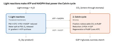
The two stages of photosynthesis are interconnected. Light reactions generate ATP and NADPH, which power the Calvin cycle. The Calvin cycle uses CO2 to produce sugars.
Light reactions (thylakoid membrane): Photosystem II absorbs light, splits water, releases O2, creates H+ gradient, ATP synthase produces ATP, NADP+ is reduced to NADPH.
Calvin cycle (stroma): Rubisco fixes CO2 to RuBP, ATP and NADPH convert 3-PGA to G3P, G3P is used to make glucose, sucrose, starch; RuBP is regenerated.
Photosynthesis Labs and Applications
Elodea + phenol red: CO2 uptake raises pH, color shifts.
Floating leaf disks: O2 production causes disks to float; rate depends on light color.
Starch test: Iodine stains photosynthesized regions blue-black.
Comparison: Both photosynthesis and respiration use H+ gradients and ATP synthase.
Practice Problems
Sample Questions and Answers
Atomic structure: Fluorine (atomic number 9, mass 19): 9 protons, 10 neutrons, 9 electrons.
Radioactive dating: ⅛ C-14 left = 3 half-lives = 17,190 years.
Experimental design: Independent variable: hours of light; dependent: plant height; control: 0-hour group.
Conversions: 250 mL = 0.25 L; 0.5 g = 500 mg; 1,500 µL = 1.5 mL.
Density: 12 g rock, 4 mL displacement: density = 3.0 g/mL.
Microscopy: 40× objective × 10× ocular = 400×; use fine focus only.
Macromolecule tests: Benedict's red, iodine yellow-brown, Biuret violet = reducing sugar and protein, no starch.
Starch detection: Benedict's cannot detect starch directly; must be hydrolyzed.
Protein secretion pathway: Nucleus → ribosome (rough ER) → vesicle → Golgi → secretory vesicle → plasma membrane.
Cell identification: No nucleus, cell wall, 70S ribosomes, 2 µm = prokaryote.
Absorption vs. action spectrum: Accessory pigments broaden action spectrum.
Calvin cycle: 6 CO2 → 12 G3P; 2 leave, 10 regenerate 6 RuBP; 2 G3P → 1 glucose.
Osmosis: Red blood cell in distilled water swells and may burst (hypotonic).
Photosynthesis lab: Green light is reflected, so disks float slower than under red light.
