 뒤로
The Molecular Basis of Inheritance: DNA Structure, Replication, and Key Experiments
스터디 가이드 - 스마트 노트
 자료에 맞춘 맞춤형 노트, 핵심 정의, 예시, 맥락을 확장해 제공합니다.
Griffith's Experiment and Bacterial Transformation
Discovery of Genetic Material
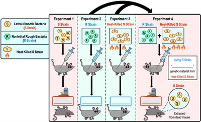
Frederick Griffith's 1928 experiment was pivotal in identifying that an unknown genetic "factor" controls the traits of organisms. Griffith demonstrated that bacteria can acquire new traits through a process called transformation, where external DNA is taken up, resulting in genotypic and phenotypic changes.
Transformation: The uptake of external DNA by a cell, leading to a change in its genotype and phenotype.
R and S Strains: The "R" (rough) strain forms rough colonies and is non-lethal, while the "S" (smooth) strain forms smooth colonies and is lethal.
Key Finding: Mixing heat-killed S strain with living R strain resulted in the transformation of R strain into lethal S strain, indicating transfer of genetic material.
Example: Griffith's experiment showed that bacteria can transform genetic material from dead cells to living cells.
The Hershey-Chase Experiment
Confirmation of DNA as Genetic Material
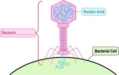
In 1952, Hershey and Chase used bacteriophages (viruses that infect bacteria) to confirm that DNA, not protein, is the genetic material. Bacteriophages consist of a protein coat surrounding nucleic acid (DNA).
Bacteriophage: A virus that infects bacteria and replicates by hijacking the bacterial machinery.
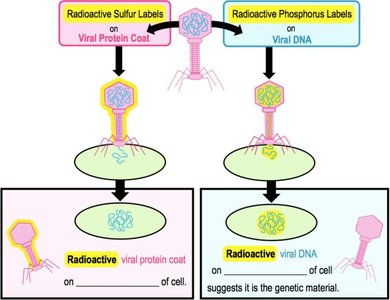
Key Finding: Only viral DNA enters the bacterial cell during infection, not the protein coat.
Experimental Design: Radioactive sulfur labeled the protein coat, while radioactive phosphorus labeled the DNA. Only radioactive DNA was found inside the bacteria, confirming DNA as the genetic material.
Example: The Hershey-Chase experiment demonstrated that DNA, not protein, is responsible for inheritance.
Chargaff's Rules
DNA Base Composition and Pairing
Erwin Chargaff made two important discoveries about DNA in the early 1950s:
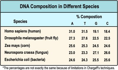
Species Variation: DNA base composition varies between species.
Base Pairing: In each species, the percentage of adenine (A) is roughly equal to thymine (T), and guanine (G) is roughly equal to cytosine (C).
Example: Chargaff's data showed consistent base pairing ratios across species, supporting the double helix model.
Species | A (%) | T (%) | G (%) | C (%) |
|---|---|---|---|---|
Homo sapiens (human) | 31.0 | 31.5 | 19.1 | 18.4 |
Drosophila melanogaster (fruit fly) | 27.3 | 27.6 | 22.5 | 22.5 |
Zea mays (corn) | 25.6 | 25.3 | 24.5 | 24.6 |
Neurospora crassa (fungus) | 23.0 | 23.3 | 27.1 | 26.6 |
Escherichia coli (bacteria) | 24.6 | 24.3 | 25.5 | 25.6 |
Discovering the Structure of DNA
X-Ray Diffraction and the Double Helix
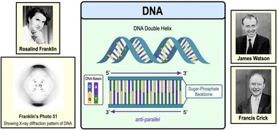
Rosalind Franklin used X-ray diffraction to capture the famous Photo 51, which provided crucial evidence for the structure of DNA. In 1953, James Watson and Francis Crick used Franklin's data to describe DNA as a double helix with two anti-parallel strands of nucleotides.
Double Helix: DNA consists of two strands twisted around each other.
Anti-parallel Strands: The two strands run in opposite directions (5' to 3' and 3' to 5').
Base Pairing: Nucleotides pair via hydrogen bonds (A–T, C–G).
Example: Watson and Crick's model explained how DNA could replicate and store genetic information.
Detailed DNA Structure
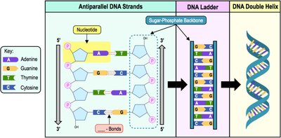
DNA is composed of nucleotide monomers linked together. Each strand has a free phosphate group at the 5' end and a free hydroxyl group (-OH) at the 3' end. The strands are held together by hydrogen bonds between complementary bases.
Nucleotide: Consists of a sugar, phosphate, and nitrogenous base.
Phosphodiester Bond: Links the phosphate group of one nucleotide to the 3' OH of the next.
Complementary Base Pairing: Ensures accurate replication and transcription.
Meselson-Stahl Experiment: DNA Replication Models
Semi-Conservative Replication
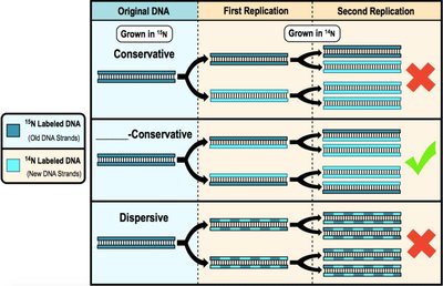
In 1958, Meselson and Stahl demonstrated that DNA replication follows the semi-conservative model. Each new DNA molecule consists of one old (parental) strand and one newly synthesized strand.
Semi-Conservative Model: Parental strands separate and serve as templates for new strands.
Experimental Evidence: Density labeling showed that after replication, DNA molecules contained one old and one new strand.
Example: The experiment confirmed that DNA replication preserves genetic information across generations.
Introduction to DNA Replication
Prokaryotic vs. Eukaryotic DNA Replication
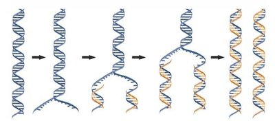
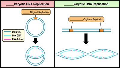
Much more is known about prokaryotic DNA replication, but the fundamental processes are similar in both prokaryotes and eukaryotes. Parental strands act as templates for synthesizing new complementary DNA.
Origin of Replication and Replication Forks
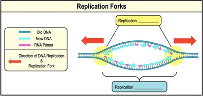
DNA replication begins at specific sequences called the origin of replication (ORI). Prokaryotes typically have one ORI per chromosome, while eukaryotes have multiple ORIs. Proteins bind to the ORI, separating the DNA strands and forming replication forks—Y-shaped regions where DNA is unwound and replication proceeds bidirectionally.
Unwinding the DNA: Topoisomerase, Helicase, and SSBs
Enzymes Involved in DNA Unwinding
Several proteins participate in the unwinding of DNA during replication:
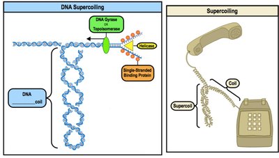
Topoisomerase (DNA Gyrase): Relieves strain caused by supercoiling ahead of the replication fork.
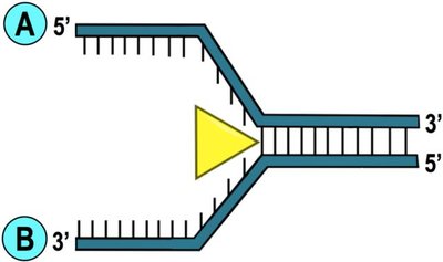
Helicase: Unwinds DNA by breaking hydrogen bonds, creating single-stranded DNA.
Single-Strand Binding Proteins (SSBs): Prevent reannealing and degradation of separated DNA strands.
DNA Polymerases and Replication Requirements
DNA Polymerase Function and Directionality
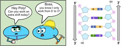
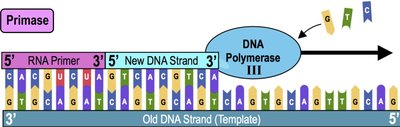
DNA polymerases are the primary enzymes responsible for building new DNA strands. New DNA is always synthesized in the 5' to 3' direction, elongating from the free 3' OH group.
Template: The parental DNA strand acts as a guide for building new strands.
Primer: A small RNA molecule provides a starting point for DNA polymerase.
Primase: Enzyme that builds RNA primers.
DNA Polymerase III: Main enzyme for elongating new DNA strands in prokaryotes.
DNA Polymerase I: Removes RNA primers and replaces them with DNA nucleotides.
Leading and Lagging DNA Strands
Mechanisms of DNA Synthesis
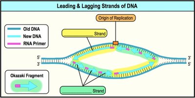
Upon separation of DNA at the replication fork, two single-stranded DNA molecules are formed:
Leading Strand: Synthesized continuously in the same direction as the replication fork movement; requires only one RNA primer.
Lagging Strand: Synthesized discontinuously in the opposite direction as the replication fork movement; replicated in small segments called Okazaki fragments, each requiring an RNA primer. Okazaki fragments are joined by DNA ligase.
Replication Fork Close-Up
DNA polymerase can only add nucleotides to the 3' end, resulting in antiparallel synthesis of the two strands.
Steps of DNA Replication
Summary of Replication Steps
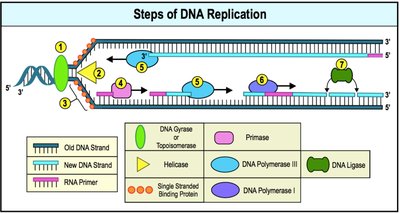
DNA replication in prokaryotes can be simplified into several steps:
Topoisomerase binds to the ORI and relieves strain due to supercoiling.
Helicase binds and unwinds the two strands of template DNA by breaking hydrogen bonds.
Single-Stranded-Binding proteins bind to single-stranded DNA.
Primase adds RNA primers to the template DNA.
DNA Polymerase III adds nucleotides to the 3' end of primers and continues elongation.
DNA Polymerase I removes RNA primers and replaces them with DNA nucleotides.
DNA Ligase covalently joins Okazaki fragments together on the lagging strand.
DNA Repair and Proofreading
Error Correction in DNA Replication
DNA replication is not always perfect; base-pairing errors can occur. Unrepaired errors result in permanent mutations, which can lead to diseases like cancer. DNA polymerases have proofreading abilities, reducing the error rate significantly. Other DNA repair enzymes correct errors not fixed by proofreading.
Telomeres and Telomerase
Chromosome End Protection and Cellular Aging
Telomeres are non-coding DNA sequences at the ends of eukaryotic chromosomes. Telomeres shorten with each round of DNA replication, which is linked to cellular aging. Significant telomere loss signals cell division to stop in normal cells. Telomerase is an enzyme found in some cells that catalyzes the lengthening of telomeres, usually expressed in germ cells and cancer cells, allowing them to maintain telomere length.
Example: Telomerase activity can extend the lifespan of human cells by preventing telomere shortening.
Summary Table: Key Enzymes in DNA Replication
Enzyme / Protein | Function |
|---|---|
Topoisomerase | Relieves DNA supercoiling ahead of replication fork |
Helicase | Unwinds DNA double helix at the replication fork |
Single-Stranded Binding Protein | Binds to and stabilizes single-stranded DNA |
Primase | Creates RNA primers as starting points for DNA synthesis |
DNA Polymerase III | Builds new DNA strand using old DNA strand as template |
DNA Polymerase I | Replaces RNA primers with DNA |
DNA Ligase | Covalently joins Okazaki fragments in lagging strand |
Additional info:
DNA replication is fundamental to inheritance and cell division.
Mutations in replication or repair enzymes can lead to genetic diseases.
Telomere research is important for understanding aging and cancer biology.
