 뒤로
뒤로The Skeletal System: Structure, Function, and Development
스터디 가이드 - 스마트 노트
 자료에 맞춘 맞춤형 노트, 핵심 정의, 예시, 맥락을 확장해 제공합니다.
The Skeletal System
Overview and Functions
The skeletal system is a dynamic organ system composed primarily of osseous (bone) tissue and additional supporting connective tissues. It is constantly remodeled through processes of bone formation and resorption. The skeletal system serves several essential functions:
Support and Movement: Provides a rigid framework for muscle attachment, enabling movement through levers and joints.
Protection: Encloses and protects vital organs such as the brain, heart, lungs, and spinal cord.
Storage and Balance: Acts as a reservoir for minerals (calcium, phosphate), electrolytes, and participates in acid-base balance.
Hematopoiesis: Site of new blood cell production in red marrow.
Energy Storage: Stores triglycerides in yellow marrow.
Hormone Production: Produces osteocalcin, which influences energy metabolism.
Classification and Structure of Bones
Bone Shapes
Bones are classified by shape, which relates to their function and location:
Long Bones: Longer than wide (e.g., humerus, femur); primarily in limbs.
Short Bones: Cube-shaped (e.g., wrist, ankle); includes sesamoid bones within tendons.
Flat Bones: Thin, flat, and slightly curved (e.g., sternum, scapulae, ribs, skull bones).
Irregular Bones: Complex shapes (e.g., vertebrae, os coxae).
Gross Structure of Bones
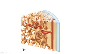
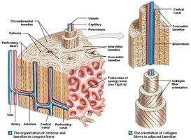
All bones consist of a compact bone layer sandwiching spongy bone. The compact (cortical) bone is organized into osteons, while spongy (trabecular) bone forms a latticework of trabeculae.
Microscopic Structure
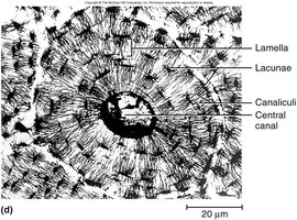
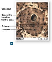
The functional unit of compact bone is the osteon, consisting of concentric lamellae surrounding a central canal. Spongy bone lacks osteons but contains trabeculae aligned along lines of stress.
Bone Tissue Composition
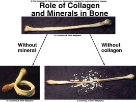
Organic Matrix (Osteoid): Produced by osteoblasts; contains collagen fibers and ground substance (GAGs) for flexibility and tensile strength.
Inorganic Matrix: Mainly hydroxyapatite (calcium phosphate crystals) for hardness and resistance to compression.
Bone Cells
Types of Bone Cells
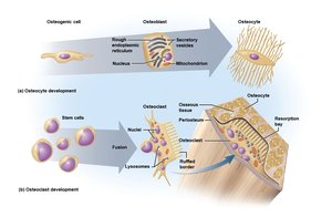
Osteogenic Cells: Stem cells in periosteum and endosteum; give rise to osteoblasts.
Osteoblasts: Bone-forming cells; synthesize osteoid and initiate calcification.
Osteocytes: Mature bone cells trapped in lacunae; maintain bone matrix and sense mechanical stress.
Osteoclasts: Large, multinucleate cells derived from hematopoietic stem cells; responsible for bone resorption.
Bone Lining Cells: Flat cells on bone surfaces; help maintain bone matrix.
Bone Development and Growth
Ossification (Osteogenesis)
Bone formation occurs through two main processes:
Intramembranous Ossification: Forms flat bones (e.g., cranial bones, clavicle) directly from mesenchymal tissue.
Endochondral Ossification: Forms most bones (long, short, irregular) by replacing a cartilage model with bone.
Endochondral Ossification Steps
Bone collar forms around diaphysis of hyaline cartilage model.
Primary ossification center develops; cartilage in center calcifies and dies.
Periosteal bud invades, forming spongy bone.
Diaphysis elongates; medullary cavity forms.
Secondary ossification centers form in epiphyses.
Epiphyses ossify; hyaline cartilage remains at articular surfaces and epiphyseal plates.

Bone Growth in Length
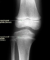
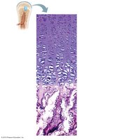
Occurs at the epiphyseal plate through interstitial growth of cartilage, followed by ossification. The plate consists of several zones:
Resting Zone: Quiescent cartilage.
Proliferation Zone: Rapid mitosis of chondrocytes.
Hypertrophic Zone: Enlargement and erosion of older cartilage cells.
Calcification Zone: Matrix calcifies, chondrocytes die, blood vessels invade.
Ossification Zone: Osteoblasts lay down new bone on spicules.
Bone Growth in Width (Appositional Growth)
Bones increase in diameter by appositional growth, where new bone is deposited by osteoblasts in the periosteum, and osteoclasts widen the medullary cavity.
Bone Remodeling and Repair
Bone Remodeling
Bone is continuously renewed through the coordinated actions of osteoclasts (resorption) and osteoblasts (deposition). This process adapts bone structure to mechanical stress and repairs microdamage.
Remodeling Rate: The entire skeleton is replaced every 5–10 years in young adults; high-stress areas remodel more frequently.
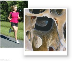
Mechanical Stress: Weight-bearing exercise stimulates bone thickening and strength.
Bone Repair
Bone repair after fracture involves hematoma formation, fibrocartilaginous callus formation, bony callus formation, and bone remodeling.
Calcium Homeostasis and Hormonal Regulation
Calcium Balance
Maintaining blood calcium levels (9–11 mg/100 mL) is critical for nerve impulse transmission and muscle contraction. Three main hormones regulate calcium homeostasis:
Parathyroid Hormone (PTH): Increases blood calcium by stimulating osteoclast activity and enhancing calcium reabsorption in kidneys.
Calcitonin: Lowers blood calcium by inhibiting osteoclasts (minor effect in adults).
Calcitriol (Active Vitamin D): Increases calcium absorption from the intestine.
Calcium is deposited in bone as hydroxyapatite and released as needed to maintain homeostasis.
Cartilage in the Skeletal System
Types and Growth of Cartilage
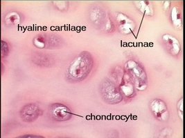
Hyaline Cartilage: Most abundant; found at articular surfaces, epiphyseal plates, and respiratory structures.
Elastic Cartilage: Flexible; found in ear and epiglottis.
Fibrocartilage: Strong; found in intervertebral discs and menisci.
Cartilage grows by two mechanisms:
Appositional Growth: New matrix added to the surface by chondroblasts in the perichondrium.
Interstitial Growth: Chondrocytes divide and secrete new matrix from within.
Clinical Considerations
Bone Disorders
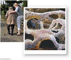
Osteoporosis: Increased bone resorption leads to porous, fragile bones; common in elderly.
Rickets: Defective mineralization in children due to vitamin D deficiency; results in bowed legs.
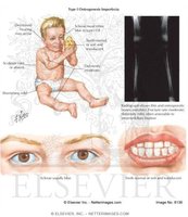
Osteogenesis Imperfecta: Genetic disorder causing brittle bones due to defective collagen.
Summary Table: Bone Cell Types and Functions
Cell Type | Origin | Function |
|---|---|---|
Osteogenic Cell | Mesenchyme | Stem cell; differentiates into osteoblasts |
Osteoblast | Osteogenic cell | Bone formation; secretes osteoid |
Osteocyte | Osteoblast | Maintains bone matrix; mechanosensor |
Osteoclast | Hematopoietic stem cell | Bone resorption; releases calcium |
Bone Lining Cell | Osteoblast | Maintains bone surface |
Additional info: This guide integrates foundational concepts from the structure and function of the skeletal system, bone development, and clinical relevance, suitable for General Biology students preparing for exams.
