 뒤로
Altered Immunity and Immune Response: Hypersensitivity, Autoimmunity, and Immunodeficiency
스터디 가이드 - 스마트 노트
 자료에 맞춘 맞춤형 노트, 핵심 정의, 예시, 맥락을 확장해 제공합니다.
Immune System Hypersensitivity and Altered Immunity
Overview of Immune Response
The immune response (IR) is a complex system that protects the body from pathogens such as bacteria and viruses, and also plays a role in detecting and eliminating cancer cells. The IR involves both cell-mediated immunity (CMI) and humoral immunity (HI), utilizing T helper (Th) cells, cytotoxic T (Tc) cells, antibodies (Ab), and memory cells for secondary responses.
Cell-mediated immunity (CMI): Involves Th and Tc cells targeting infected or abnormal cells.
Humoral immunity (HI): Involves B cells producing antibodies that neutralize pathogens.
Key functions: Opsonization, neutralization, precipitation, agglutination, complex formation, and cell lysis.
Altered Immune Response
Alterations in the immune response can lead to disease, including hypersensitivity (allergy), autoimmunity, and immune deficiency (e.g., AIDS).
Hypersensitivity: Excessive or inappropriate immune response to antigens.
Autoimmunity: Immune response directed against self-antigens.
Immunodeficiency: Reduced or absent immune response.
Types of Hypersensitivity Reactions
Classification of Hypersensitivity
Hypersensitivity reactions are classified into four types based on the immune mechanisms involved:
Type I: IgE antibody-mediated, no complement (C') involvement.
Type II: IgM or IgG antibody-mediated, complement involved, cell/tissue targeted.
Type III: IgM or IgG antibody-mediated, complement involved, immune complexes precipitate in tissues.
Type IV: T cell-mediated (Tc), delayed-type, no antibody or complement involved.
Type I: Allergy and Anaphylaxis
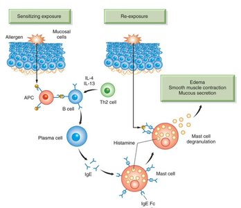
Type I hypersensitivity is characterized by IgE-mediated reactions to environmental allergens, leading to allergic responses and potentially anaphylaxis.
Genetic predisposition: Atopic individuals produce IgE to specific allergens.
First exposure: IgE is produced and binds to mast cells/basophils.
Re-exposure: Allergen binds to IgE, causing degranulation and histamine (H1) release.
Complications: Local or systemic symptoms depending on tissue involvement.
Signs and Symptoms of Allergy and Histamine Action
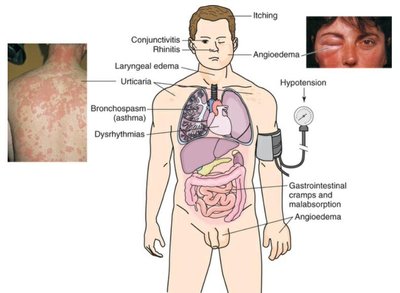
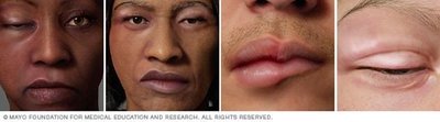
Local effects: Angioedema, urticaria, redness, warmth, pruritus, mucous production.
Systemic effects: Widespread edema, hypotension, pulmonary edema, bronchoconstriction, vomiting.
Diagnosis and Treatment of Allergy
Diagnosis: Based on signs and symptoms, allergy testing, serum IgE levels, oral food challenge.
Prevention: Avoidance of allergen.
Treatment: Desensitization shots, antihistamines, epinephrine for anaphylaxis, OIT/SLIT.
Allergy Testing
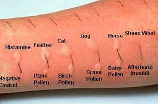
Skin prick tests are used to identify specific allergens by observing local reactions.

Examples of Allergy
Hay fever, allergic asthma, food allergy, bee/wasp sting, latex allergy, drug allergy, environmental allergy.
Key Issues in Allergy and Medication Administration
Allergy is IgE-mediated; first exposure is required for IgE production.
Cross-sensitivity can occur with similar antigens (e.g., penicillins and cephalosporins).
Distinguish between true allergy and side effects (e.g., codeine-induced constipation).
Anaphylaxis
Definition: Severe, life-threatening allergic reaction.
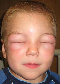
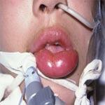
Symptoms: Difficulty swallowing/breathing, vasodilation, angioedema, hypotension.

Early Signs and Symptoms of Anaphylaxis
Warm, flushed skin, systemic angioedema, urticaria, bronchial/laryngeal/tracheal spasm, pruritus, low BP, GI upset.
Compensatory Mechanisms in Anaphylactic Shock
Fluid shift, HPA axis, RAA, ADH activation: decreased BP, increased HR, cool pale skin, decreased urine output, increased RR, altered LOC, peripheral edema, bronchospasm.
Treatment of Anaphylaxis
Epinephrine (IM), stop exposure, airway management, IV fluids, oxygen, antihistamines, steroids, bronchodilators, H2 receptor antagonists.
Type II and III Hypersensitivity Reactions
Type II: Cell and Tissue-Specific, Antibody-Mediated
Type II reactions target specific cell or tissue antigens, leading to cell damage and inflammation.
Mechanism: IgG/IgM antibodies bind to cell/tissue antigens, activate complement, cause cell lysis and inflammation.
Examples: Grave’s disease, hemolytic disease of the newborn (HDN), blood transfusion reaction.
Type III: Immune Complex Precipitation
Type III reactions involve immune complexes forming in circulation and precipitating in tissues, causing severe inflammation and tissue damage.
Mechanism: Ag-Ab complexes activate complement, attract WBCs, degranulate basophils, cause inflammation.
Examples: Raynaud’s disease, celiac disease, HDN, blood transfusion reaction.
Signs and Symptoms of Types II and III
Inflammation, loss of tissue function, hemolysis, organ damage, more severe in Type III.
ABO and Rh Incompatibility
Blood transfusion reactions and hemolytic disease of the newborn are classic examples of Type II and III hypersensitivity.
Blood Type | Antigen (Ag) | Antibody (Ab) |
|---|---|---|
A | A | anti-B |
B | B | anti-A |
AB | A & B | none |
O | none | anti-A, anti-B |
Rh+ | Rh | none |
Rh- | none | anti-Rh |
Blood Transfusion Reaction: Type II
Recipient’s antibodies react with donor RBC antigens, activate complement, cause hemolysis, inflammation, hypotension, flushing, urticaria, hypoxia, fever, hemolytic anemia, jaundice.
Blood Transfusion Reaction: Type III
Immune complexes precipitate in capillaries, causing organ damage (kidney, lungs, joints, skin, heart), flank pain, chest/back pain, SOB, joint pain.
Complications of Type III
Excess unconjugated bilirubin causes CNS toxicity, jaundice, spleen enlargement, renal damage, organ damage.
Diagnosis and Treatment of Transfusion Reaction
Stop transfusion, diagnose by S&S, blood tests, treat with IV fluids, O2, prevent with proper ABO/Rh testing.
Hemolytic Disease of the Newborn (HDN)
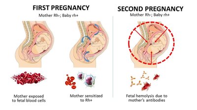
HDN is a Type II/III reaction where maternal antibodies attack fetal RBCs, leading to anemia, jaundice, and potential CNS damage.
First fetus: Rh- mother exposed to Rh+ fetal cells at delivery, forms anti-Rh antibodies.
Second fetus: Maternal anti-Rh antibodies cross placenta, destroy fetal RBCs, cause anemia, jaundice, kernicterus, stillbirth.
Diagnosis and Treatment of HDN
Diagnose by S&S, blood tests, bilirubin levels, treat with transfusion and support, prevent with Rhogam for Rh- mothers.
Type IV Hypersensitivity Reactions
Type IV: T Cell-Mediated (Delayed-Type)
Type IV reactions are mediated by cytotoxic T cells, with no antibody involvement. They appear several days post-exposure and cause tissue destruction.
Examples: Graft vs host (transplant rejection), Type I diabetes mellitus, contact dermatitis, vaccine/drug reactions.
Autoimmunity
Mechanisms and Examples of Autoimmune Diseases
Autoimmunity occurs when the immune system produces antibodies against self-antigens, leading to tissue destruction and chronic inflammation.
Genetic factors: Associated with certain genes/diseases.
Breakdown in tolerance: Self-antigens are targeted as foreign.
Examples: Inflammatory bowel disease, rheumatoid arthritis, systemic lupus erythematosus (SLE), ankylosing spondylitis, myasthenia gravis, multiple sclerosis, psoriasis.
Pathophysiology of Autoimmune Diseases
Auto-antibody formation, Ag-auto-Ab complex, inflammation, complement activation, tissue destruction, loss of function, pain, scar formation.
Immunodeficiency: HIV and AIDS
Transmission and Pathophysiology of HIV
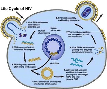
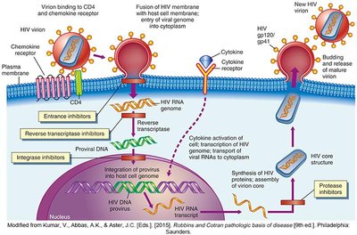
HIV is a virus transmitted via blood and body fluids, leading to acquired immune deficiency syndrome (AIDS) if untreated. It targets CD4 receptors on Th cells, integrates into host genome, and destroys immune cells.
Transmission: Sexual activity, IV needle sharing, maternal/child transmission, lack of precautions.
Life cycle: Attachment to CD4, entry, reverse transcription, integration, replication, cell lysis, spread to other cells.
Effects of HIV on Immune System
Loss of Th cells, B cells, lymphocytes, macrophages; loss of immunity and inflammation; susceptibility to infections.
Organ-specific effects: GI tract (malnutrition), skin (Kaposi’s sarcoma), brain (dementia).
Spectrum of HIV Disease
Initial infection: mild, flu-like symptoms.
Immunosuppression: CD4 count < 500.
Opportunistic infections: CD4 count < 200 (e.g., candidiasis, TB, herpes simplex, pneumocystis jiroveci pneumonia, varicella zoster).
Immune Response to HIV
Antibodies produced are not protective; loss of CMI due to Th cell depletion allows viral spread.
Diagnosis and Treatment of HIV
Blood test for HIV antibodies (seropositive), viral load monitoring, multiple drug therapy (not curative), window period ~6 months.
HIV Vaccine and Prevention
Vaccine in clinical trials; difficulty due to antigenic variation.
Prevention: safer behaviors, PPE, avoid needle stick injury, PReP and PEP protocols.
Conclusion
The immune response is essential for protection against infectious agents, but alterations can lead to hypersensitivity, autoimmunity, or immunodeficiency. Understanding these mechanisms is crucial for diagnosis, treatment, and prevention of related diseases.
