 뒤로
The Innate Immune System: Structure and Function
스터디 가이드 - 스마트 노트
 자료에 맞춘 맞춤형 노트, 핵심 정의, 예시, 맥락을 확장해 제공합니다.
The Innate Immune System
Immune System Components
The immune system is composed of various cells and tissues that work together to defend the body against pathogens. The primary cellular components are leukocytes, which are divided into granulocytes and agranulocytes based on the presence of granules in their cytoplasm.
Leukocytes
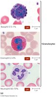
Granulocytes: Include neutrophils, eosinophils, and basophils. These cells contain cytoplasmic granules and play key roles in inflammation, phagocytosis, and defense against parasites.
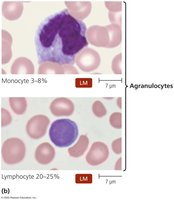
Agranulocytes: Include monocytes and lymphocytes. These cells lack visible granules and are involved in phagocytosis and adaptive immunity.
Granulocytes
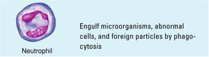
Neutrophils: Engulf microorganisms, abnormal cells, and foreign particles by phagocytosis. They are the most abundant leukocytes and are the first responders to infection.
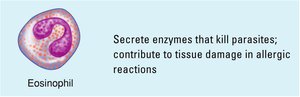
Eosinophils: Secrete enzymes that kill parasites and contribute to tissue damage in allergic reactions.
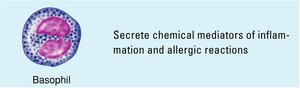
Basophils: Secrete chemical mediators of inflammation and allergic reactions, such as histamine.
Agranulocytes
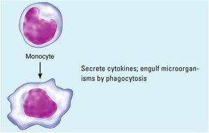
Monocytes: Circulate in the blood and differentiate into macrophages and dendritic cells in tissues. They secrete cytokines and engulf microorganisms by phagocytosis.
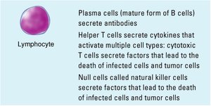
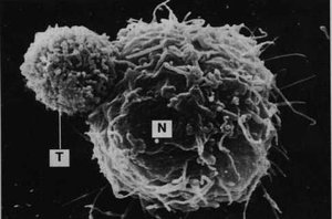
Lymphocytes: Include B cells, T cells, and natural killer (NK) cells. B cells produce antibodies, T cells mediate cellular immunity, and NK cells target infected or abnormal cells.
Dendritic Cells
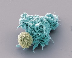
Dendritic cells are specialized antigen-presenting cells that capture antigens and present them to T cells, initiating adaptive immune responses. They are found in tissues in contact with the external environment.
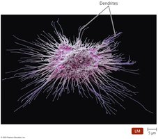
Lymphoid Tissues
Lymphoid tissues are organized structures that support the development and function of immune cells. They are classified as central (primary) or peripheral (secondary) lymphoid tissues.
Central lymphoid tissues: Bone marrow and thymus, where lymphocytes are produced and mature.
Peripheral lymphoid tissues: Spleen, lymph nodes, tonsils, adenoids, appendix, and Peyer’s patches, where immune responses are initiated.
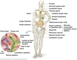
Central Lymphoid Tissue
Bone marrow: Site of hematopoiesis (formation of blood cells), including all leukocytes.
Thymus: Site of T cell maturation.
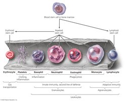
Peripheral Lymphoid Tissue
Includes spleen, lymph nodes, tonsils, adenoids, appendix, and Peyer’s patches.
These tissues trap microorganisms and foreign particles, exposing them to high concentrations of leukocytes for immune surveillance and response.
Organization of the Body’s Defenses
Innate Immunity (Nonspecific Defenses)
Innate immunity provides immediate, nonspecific defense against pathogens through physical barriers, chemical mediators, and cellular responses. It is present from birth and does not require prior exposure to pathogens.
First Line of Defense: Physical Barriers, Chemicals, and Processes
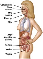
Skin: Acts as a physical barrier with constant turnover and secretions such as defensins, lysozyme, and sebum that inhibit microbial growth.
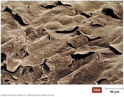
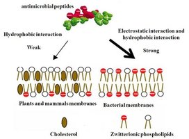
Defensins: Antimicrobial peptides that disrupt microbial membranes.
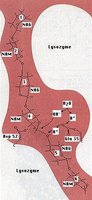
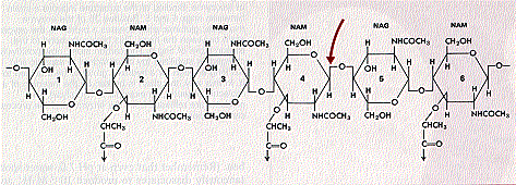
Lysozyme: Enzyme that breaks down bacterial cell walls, especially peptidoglycan.
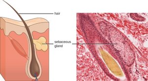
Sebum: Oily secretion from sebaceous glands that lubricates and protects the skin, and serves as a nutrient for resident microbiota.
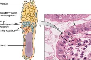
Mucous membranes: Line the respiratory, digestive, urinary, and reproductive tracts, producing mucus that traps pathogens.
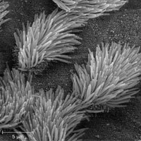
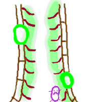
Mucociliary escalator: Cilia move mucus and trapped pathogens out of the respiratory tract.
Lacrimal Apparatus
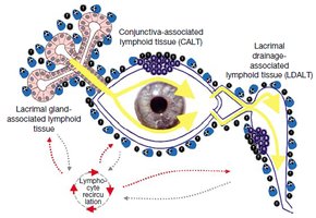
The lacrimal apparatus produces and drains tears, which contain lysozyme and lactoferrin, providing mechanical and chemical protection for the eyes.
Normal Microbiota
Normal microbiota (resident flora) are non-pathogenic microorganisms that inhabit body surfaces and compete with pathogens, providing microbial antagonism.
Other Secretions & Activities
Secretions and activities from the urinary, reproductive, and cardiovascular systems also contribute to the first line of defense by flushing out pathogens and maintaining sterile environments.
Second Line of Defense: Internal Defenses
When pathogens breach the first line of defense, the second line includes cellular and chemical responses such as phagocytosis, nonphagocytic killing, nonspecific chemical defenses, inflammation, and fever.
Phagocytosis
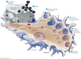
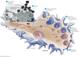
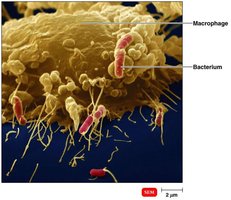
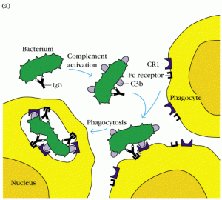
Phagocytosis is the process by which certain cells (neutrophils, macrophages, dendritic cells) engulf and destroy pathogens. The steps include chemotaxis, adherence, ingestion, fusion with lysosome, killing, and elimination.
Nonphagocytic Killing
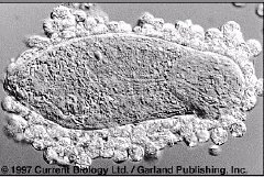
Eosinophils: Kill parasites and contribute to allergic tissue damage.
Natural Killer (NK) Cells: Destroy infected or abnormal cells by releasing cytotoxic molecules.
Nonspecific Chemical Defenses
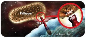
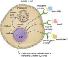
Pattern Recognition Receptors (PRRs), such as Toll-Like Receptors (TLRs) and NOD-like Receptors (NLRs), are expressed by phagocytes and recognize Pathogen-Associated Molecular Patterns (PAMPs), triggering immune responses.
Interferons
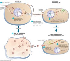
Interferons (alpha and beta) are cytokines produced by virus-infected cells that protect neighboring cells by inducing the production of antiviral proteins (AVPs).
Complement System
The complement system is a group of serum proteins that, when activated, lead to cell lysis, inflammation, and opsonization. There are three activation pathways: classical, alternative, and lectin.
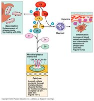
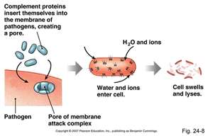
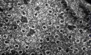
Membrane Attack Complex (MAC): Formed by complement proteins (C5b, C6, C7, C8, C9), it creates pores in pathogen membranes, leading to lysis.
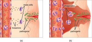
Inflammation: Complement fragments (C5a, C3a) induce mast cell degranulation and attract phagocytes.
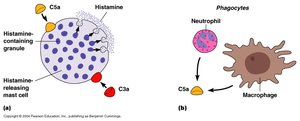
Opsonization: C3b acts as an opsonin, coating pathogens to enhance phagocytosis.
Inflammation
Inflammation is a series of events that result in the accumulation of proteins, fluid, and phagocytes at sites of injury or infection. Symptoms include redness, heat, edema, pain, and loss of function, primarily due to the effects of histamine and other mediators.
Fever
Fever is an elevation of body temperature above 37°C, induced by pyrogens. It enhances immune responses and inhibits the growth of some pathogens.
