 뒤로
뒤로Anatomy & Physiology Nervous System Study Guide
스터디 가이드 - 스마트 노트
 자료에 맞춘 맞춤형 노트, 핵심 정의, 예시, 맥락을 확장해 제공합니다.
Q1. Define the following terms related to the Central Nervous System: Neurons, Glial cells, Soma, Axon, Dendrite, Gray matter, White matter.
Background
Topic: Structure and Function of the Nervous System
This question tests your understanding of the basic cellular components and structural organization of the central nervous system (CNS).
Key Terms:
Neurons: Specialized cells that transmit electrical and chemical signals in the nervous system.
Glial cells: Supportive cells in the CNS that provide structural and metabolic support to neurons.
Soma: The cell body of a neuron, containing the nucleus and most organelles.
Axon: A long, slender projection that conducts electrical impulses away from the soma.
Dendrite: Branch-like extensions that receive signals from other neurons.
Gray matter: Regions of the CNS containing neuron cell bodies and dendrites.
White matter: Regions of the CNS composed mainly of myelinated axons.
Step-by-Step Guidance
Start by identifying the main cell types in the CNS: neurons and glial cells.
Describe the structure of a neuron, including the soma, axon, and dendrite.
Explain the functional differences between gray matter and white matter in the CNS.
Consider how glial cells support neurons and contribute to CNS function.
Try solving on your own before revealing the answer!
Final Answer:
Neurons: Cells specialized for transmitting electrical impulses and processing information.
Glial cells: Support cells that maintain homeostasis, form myelin, and provide support and protection for neurons.
Soma: The cell body of a neuron, where most metabolic activity occurs.
Axon: The part of the neuron that carries signals away from the soma toward other cells.
Dendrite: Extensions from the soma that receive incoming signals.
Gray matter: Contains neuron cell bodies, dendrites, and unmyelinated axons; involved in processing information.
White matter: Consists mainly of myelinated axons; responsible for transmitting signals between different parts of the CNS.
These terms are fundamental to understanding the structure and function of the nervous system.
Q2. Define the following terms related to the Spinal Cord: Ganglion vs. nucleus, Tract.
Background
Topic: Organization of the Spinal Cord
This question focuses on the anatomical terminology used to describe clusters of neurons and pathways in the spinal cord.
Key Terms:
Ganglion: A cluster of neuron cell bodies located outside the CNS.
Nucleus: A cluster of neuron cell bodies located within the CNS.
Tract: A bundle of axons within the CNS that carry information.
Step-by-Step Guidance
Compare the location and function of ganglia and nuclei.
Describe what a tract is and how it relates to the transmission of information in the spinal cord.
Think about how these structures contribute to the overall function of the nervous system.
Try solving on your own before revealing the answer!
Final Answer:
Ganglion: Cluster of neuron cell bodies outside the CNS (e.g., dorsal root ganglion).
Nucleus: Cluster of neuron cell bodies within the CNS (e.g., thalamic nuclei).
Tract: Bundle of axons in the CNS that carry signals between regions.
Understanding these terms helps clarify the organization of neural pathways.
Q3. Define Afferent and Efferent neurons in the Peripheral Nervous System.
Background
Topic: Peripheral Nervous System Function
This question tests your knowledge of the direction of information flow in the nervous system.
Key Terms:
Afferent neurons: Carry sensory information toward the CNS.
Efferent neurons: Carry motor commands away from the CNS to effectors.
Step-by-Step Guidance
Identify the direction of information flow for afferent and efferent neurons.
Relate these terms to sensory and motor functions.
Consider examples of each type of neuron in the PNS.
Try solving on your own before revealing the answer!
Final Answer:
Afferent neurons: Transmit sensory information from the body to the CNS.
Efferent neurons: Transmit motor commands from the CNS to muscles and glands.
These terms are essential for understanding how the nervous system communicates with the body.
Q4. What are the three main functional divisions of the nervous system: Sensation, Response, and Integration?
Background
Topic: Functional Organization of the Nervous System
This question tests your understanding of how the nervous system processes information and coordinates actions.
Key Terms:
Sensation: Detection of stimuli by sensory receptors.
Response: Activation of effectors (muscles or glands) based on sensory input.
Integration: Processing and interpretation of sensory information in the CNS.
Step-by-Step Guidance
Define each functional division: sensation, response, and integration.
Explain how these functions interact to produce coordinated behavior.
Provide examples of each function in the nervous system.
Try solving on your own before revealing the answer!
Final Answer:
Sensation: The nervous system detects changes in the environment.
Response: The nervous system activates effectors to produce a reaction.
Integration: The CNS processes and interprets sensory input to determine the appropriate response.
These divisions are fundamental to understanding nervous system function.
Q5. What is the role of the Autonomic Nervous System (ANS)?
Background
Topic: Autonomic Nervous System Function
This question tests your understanding of involuntary control of body functions.
Key Terms:
ANS: Controls involuntary functions such as heart rate, digestion, and glandular activity.
Enteric Nervous System: Regulates the digestive tract.
Step-by-Step Guidance
Identify the main effectors controlled by the ANS: smooth muscle, cardiac muscle, and glands.
Describe the role of the enteric nervous system within the ANS.
Explain how the ANS maintains homeostasis.
Try solving on your own before revealing the answer!
Final Answer:
ANS: Regulates involuntary functions such as heart rate, digestion, respiratory rate, and glandular secretion.
Enteric Nervous System: Controls the function of the digestive tract independently of the CNS.
The ANS is essential for maintaining internal balance and responding to stress.
Q6. Define the following terms related to nervous tissue: Synapse, Axon hillock, Node of Ranvier, Axon terminal, Synaptic end bulb.
Background
Topic: Structure of Nervous Tissue
This question tests your knowledge of the specialized structures involved in neural communication.
Key Terms:
Synapse: Junction where neurons communicate with each other or with effectors.
Axon hillock: Region where the axon originates from the soma; site of action potential initiation.
Node of Ranvier: Gaps in the myelin sheath that facilitate rapid signal transmission.
Axon terminal: End of the axon where neurotransmitters are released.
Synaptic end bulb: Swelling at the axon terminal containing neurotransmitter vesicles.
Step-by-Step Guidance
Define each term and describe its role in neural communication.
Explain how the axon hillock and node of Ranvier contribute to action potential propagation.
Describe the process of neurotransmitter release at the axon terminal and synaptic end bulb.
Try solving on your own before revealing the answer!
Final Answer:
Synapse: The site of communication between neurons or between a neuron and an effector cell.
Axon hillock: The region where the axon emerges from the soma and where action potentials are initiated.
Node of Ranvier: Gaps in the myelin sheath that allow for saltatory conduction of action potentials.
Axon terminal: The distal end of the axon where neurotransmitters are released.
Synaptic end bulb: The enlarged tip of the axon terminal containing vesicles filled with neurotransmitters.
These structures are critical for efficient communication in the nervous system.
Q7. What are the types of neurons according to axon shape: Unipolar, Bipolar, Multipolar?
Background
Topic: Neuron Classification
This question tests your understanding of the structural diversity of neurons.
Key Terms:
Unipolar: Neurons with a single process extending from the soma.
Bipolar: Neurons with two processes (one axon, one dendrite).
Multipolar: Neurons with multiple processes (one axon, many dendrites).
Step-by-Step Guidance
Define each neuron type based on the number and arrangement of processes.
Provide examples of where each type is found in the body.
Consider how structure relates to function for each neuron type.
Try solving on your own before revealing the answer!
Final Answer:
Unipolar: One process; common in sensory neurons.
Bipolar: Two processes; found in the retina and olfactory epithelium.
Multipolar: Multiple processes; most common type in the CNS.
Neuron shape is closely related to their function and location.
Q8. What are the types of glial cells and their functions?
Background
Topic: Glial Cell Diversity
This question tests your knowledge of the supportive cells in the CNS and PNS.
Key Terms:
Astrocyte: Maintains the blood-brain barrier.
Oligodendrocyte: Forms myelin in the CNS.
Microglia: Immune cells of the CNS.
Ependymal cell: Produces cerebrospinal fluid.
Satellite cell: Supports neurons in the PNS.
Schwann cell: Forms myelin in the PNS.
Step-by-Step Guidance
List the main glial cell types in the CNS and PNS.
Describe the function of each glial cell.
Explain how glial cells contribute to nervous system health and function.
Try solving on your own before revealing the answer!
Final Answer:
Astrocyte: Maintains the blood-brain barrier and supports neurons.
Oligodendrocyte: Produces myelin in the CNS.
Microglia: Acts as immune cells in the CNS.
Ependymal cell: Produces cerebrospinal fluid and lines ventricles.
Satellite cell: Supports neurons in the PNS.
Schwann cell: Produces myelin in the PNS.
Glial cells are essential for maintaining the environment and function of neurons.
Q9. What is the function of the nervous system: Thermoreceptor, Graded Potential, Threshold, Action Potential, Neurotransmitter, Thalamus, Cerebral cortex, Upper Motor Neuron, Precentral Gyrus of the Frontal Cortex, Lower Motor Neuron?
Background
Topic: Nervous System Function and Structure
This question tests your understanding of sensory detection, signal transmission, and motor control.
Key Terms:
Thermoreceptor: Detects temperature changes.
Graded Potential: Local changes in membrane potential.
Threshold: Minimum stimulus required to trigger an action potential.
Action Potential: Rapid electrical signal transmitted by neurons.
Neurotransmitter: Chemical messenger released at synapses.
Thalamus: Relay station for sensory information.
Cerebral cortex: Site of higher brain functions.
Upper Motor Neuron: Initiates voluntary movement.
Precentral Gyrus: Primary motor cortex.
Lower Motor Neuron: Directly innervates skeletal muscle.
Step-by-Step Guidance
Define each term and its role in nervous system function.
Explain how sensory detection leads to graded potentials and action potentials.
Describe the pathway from the cerebral cortex to motor neurons.
Try solving on your own before revealing the answer!
Final Answer:
Thermoreceptor: Detects temperature changes.
Graded Potential: Localized change in membrane potential.
Threshold: Minimum level needed to trigger an action potential.
Action Potential: All-or-none electrical signal.
Neurotransmitter: Chemical messenger at synapses.
Thalamus: Sensory relay center.
Cerebral cortex: Higher-order processing.
Upper Motor Neuron: Initiates movement.
Precentral Gyrus: Primary motor area.
Lower Motor Neuron: Executes movement.
These terms describe the flow of information from sensation to response.
Q10. What are the types of channels in cell membranes: Non-specific channel, Gated channel (Ligand-gated, Ionotropic, Mechanically gated, Voltage-gated, Leakage channel)?
Background
Topic: Membrane Channels and Signal Transmission
This question tests your understanding of how ions move across cell membranes and how channels are regulated.
Key Terms:
Non-specific channel: Allows passage of multiple ion types.
Gated channel: Opens or closes in response to specific stimuli.
Ligand-gated channel: Opens when a signaling molecule binds.
Ionotropic receptor: Directly controls ion flow.
Mechanically gated channel: Opens in response to physical deformation.
Voltage-gated channel: Opens in response to changes in membrane potential.
Leakage channel: Always open, allowing ions to passively diffuse.
Step-by-Step Guidance
List the types of channels and describe their mechanisms of opening and closing.
Explain the role of each channel type in generating and propagating electrical signals.
Relate channel function to nervous system activity.
Try solving on your own before revealing the answer!
Final Answer:
Non-specific channel: Permits passage of multiple ions.
Gated channel: Opens/closes in response to stimuli.
Ligand-gated channel: Opens when a ligand binds.
Ionotropic receptor: Directly controls ion flow.
Mechanically gated channel: Opens with physical force.
Voltage-gated channel: Opens with changes in voltage.
Leakage channel: Always open, allows passive ion movement.
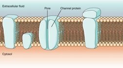
The diagram shows a channel protein forming a pore in the membrane, illustrating how ions can move across the cell membrane.
Q11. What is resting membrane potential and action potential? Define depolarization and repolarization.
Background
Topic: Membrane Potential and Neural Signaling
This question tests your understanding of the electrical properties of neurons and how signals are generated.
Key Terms and Formulas:
Resting membrane potential: The voltage difference across the membrane when the cell is at rest.
Action potential: Rapid change in membrane potential that propagates along the axon.
Depolarization: Membrane potential becomes less negative.
Repolarization: Membrane potential returns to resting value.
Key formula:
Step-by-Step Guidance
Define resting membrane potential and explain how it is maintained.
Describe the sequence of events during an action potential.
Explain the processes of depolarization and repolarization.
Try solving on your own before revealing the answer!
Final Answer:
Resting membrane potential: The stable voltage across the membrane when the neuron is not firing, typically around -70 mV.
Action potential: A rapid, temporary change in membrane potential that travels along the axon.
Depolarization: The membrane potential becomes less negative (moves toward zero).
Repolarization: The membrane potential returns to its resting value after depolarization.
These processes are essential for neural communication.
Q12. What is the difference between continuous and saltatory conduction?
Background
Topic: Action Potential Propagation
This question tests your understanding of how action potentials travel along axons.
Key Terms:
Continuous conduction: Occurs in unmyelinated axons; action potential moves along every part of the membrane.
Saltatory conduction: Occurs in myelinated axons; action potential jumps from node to node.
Step-by-Step Guidance
Define continuous and saltatory conduction.
Explain how myelin affects the speed of action potential propagation.
Describe the role of the node of Ranvier in saltatory conduction.
Try solving on your own before revealing the answer!
Final Answer:
Continuous conduction: Action potential travels along the entire length of unmyelinated axons.
Saltatory conduction: Action potential jumps between nodes of Ranvier in myelinated axons, increasing speed.
Saltatory conduction is much faster due to the presence of myelin.
Q13. What are the two types of receptors that acetylcholine can bind to?
Background
Topic: Neurotransmitter Receptors
This question tests your understanding of cholinergic signaling.
Key Terms:
Nicotinic receptor: Ionotropic receptor for acetylcholine.
Muscarinic receptor: Metabotropic receptor for acetylcholine.
Step-by-Step Guidance
Identify the two main receptor types for acetylcholine.
Describe the difference between ionotropic and metabotropic receptors.
Explain where each receptor type is found in the body.
Try solving on your own before revealing the answer!
Final Answer:
Nicotinic receptor: Ionotropic, found at neuromuscular junctions.
Muscarinic receptor: Metabotropic, found in the CNS and PNS.
Acetylcholine can bind to both types, producing different effects.
Q14. Which receptor uses a G protein: ionotropic or metabotropic?
Background
Topic: Signal Transduction
This question tests your understanding of receptor mechanisms.
Key Terms:
G protein: Intracellular protein involved in signal transduction.
Ionotropic receptor: Directly controls ion channels.
Metabotropic receptor: Indirectly controls ion channels via G proteins.
Step-by-Step Guidance
Define ionotropic and metabotropic receptors.
Explain the role of G proteins in signal transduction.
Identify which receptor type uses G proteins.
Try solving on your own before revealing the answer!
Final Answer:
Metabotropic receptors use G proteins to transmit signals inside the cell, while ionotropic receptors do not.
G protein-coupled receptors are important for many signaling pathways.
