 뒤로
뒤로Anatomy and Physiology of the Gastrointestinal Tract: Structure, Function, and Histology
스터디 가이드 - 스마트 노트
 자료에 맞춘 맞춤형 노트, 핵심 정의, 예시, 맥락을 확장해 제공합니다.
Gastrointestinal Tract: Overview and Functions
Introduction to the Gastrointestinal Tract
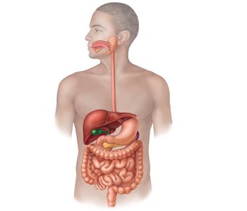
The gastrointestinal (GI) tract, also known as the alimentary canal, is a continuous muscular tube that digests and absorbs food. It extends from the mouth to the anus and includes several specialized organs and accessory structures.
Main organs: mouth, pharynx, esophagus, stomach, small intestine (duodenum, jejunum, ileum), large intestine (colon, rectum, anus)
Accessory organs: teeth, tongue, salivary glands, liver, gallbladder, pancreas
Major Functions of the GI Tract
Mastication: Mechanical and chemical breakdown of food in the mouth
Ingestion: Intake of food and swallowing
Digestion: Mechanical and chemical breakdown of food into absorbable units
Absorption: Movement of nutrients from the gut into the blood
Excretion (Defaecation): Removal of undigested material as feces
Structural Organization of the GI Tract
Layers of the GI Tract
The wall of the GI tract is composed of four main layers, each with distinct functions:
Mucosa: Innermost layer; consists of epithelium (mainly simple columnar), lamina propria (connective tissue), and muscularis mucosae (smooth muscle). Functions in secretion, absorption, and protection.
Submucosa: Dense connective tissue containing blood vessels, lymphatics, glands, and the submucosal plexus (controls secretion).
Muscularis Externa: Smooth muscle responsible for peristalsis and segmentation; contains the myenteric plexus (controls motility).
Serosa: Outermost layer; visceral peritoneum covering the GI tract in the abdominal cavity.
Movements of the GI Tract
GI tract motility is essential for moving and mixing food:
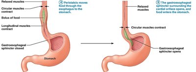
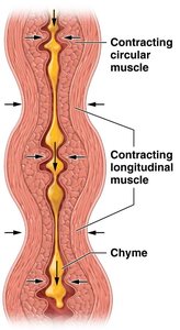
Peristalsis: Coordinated, wave-like contractions of circular and longitudinal muscles that propel food distally.
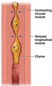
Segmentation: Alternating contractions of nonadjacent segments, mixing food and enhancing absorption.
Peritoneum and Peritoneal Folds
Structure and Function of the Peritoneum
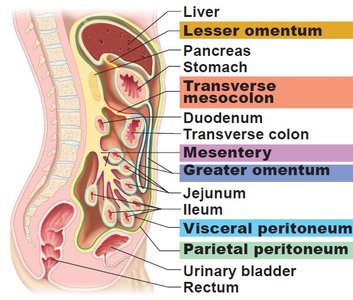
The peritoneum is a serous membrane lining the abdominal cavity and covering abdominal organs. It consists of:
Parietal peritoneum: Lines the abdominal wall
Visceral peritoneum: Covers the organs
Peritoneal cavity: Space between the two layers, containing lubricating fluid
Peritoneal Folds and Mesenteries
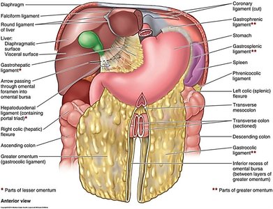
Mesentery: Double layer of peritoneum that suspends the small intestine from the posterior abdominal wall, providing a pathway for blood vessels, nerves, and lymphatics.
Mesocolon: Attaches the large intestine to the posterior abdominal wall.
Greater omentum: Large apron-like fold of visceral peritoneum that hangs from the stomach and covers the intestines, containing fat and immune cells.
Functional Anatomy of the Mouth
Oral Cavity Structure
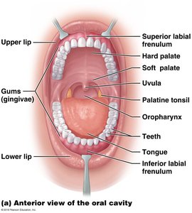
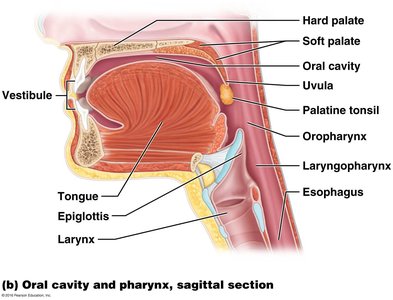
The mouth (oral cavity) is the entry point for food and is lined with stratified squamous epithelium. Major structures include:
Cheeks, hard and soft palate, uvula, tongue, teeth
Salivary Glands and Saliva
Major glands: Parotid, submandibular, sublingual
Saliva: Contains water, mucus, and the enzyme salivary amylase (initiates starch digestion)
Salivation is controlled by the parasympathetic nervous system (CN VII and IX)
Teeth: Structure and Types
Teeth are specialized for mechanical digestion (mastication):
Structure: Crown (covered by enamel), neck, root (covered by cementum), pulp cavity (contains nerves and vessels), periodontal ligaments anchor teeth to bone.
Types: Incisors (cutting), canines (tearing), premolars and molars (crushing and grinding)
Dental formula: Deciduous (20), permanent (32)
Tongue: Structure and Function
Composed of intrinsic and extrinsic muscles
Functions: Manipulates food, forms bolus, initiates swallowing, essential for speech and taste
Papillae: Filiform (friction), fungiform and vallate (taste buds)
Contains lingual tonsil (immune function)
Digestion in the Mouth
Mechanical digestion: Chewing breaks food into smaller pieces and mixes it with saliva
Chemical digestion: Salivary amylase begins starch breakdown; process called hydrolysis
Amylase is inactivated by stomach acid
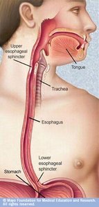
Oesophagus: Structure and Function
Anatomy of the Oesophagus
The oesophagus is a muscular tube that transports food from the pharynx to the stomach. It passes through the mediastinum and diaphragm (at the oesophageal hiatus, T10).
Four layers: mucosa, submucosa, muscle layer, outer connective tissue
Upper and lower oesophageal sphincters regulate passage of food
Peristalsis moves the bolus toward the stomach
Stomach: Structure, Histology, and Function
Gross Anatomy of the Stomach
Regions: Cardia, fundus, body, pyloric antrum, pyloric canal, pylorus (with pyloric sphincter)
Borders: Lesser curvature (medial), greater curvature (lateral)
Located under the diaphragm in the left upper abdomen
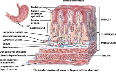
Stomach Wall and Gastric Mucosa
Mucosa: Simple columnar epithelium with rugae (folds) and gastric pits leading to gastric glands
Submucosa: Contains blood vessels and submucosal plexus
Muscularis externa: Multiple smooth muscle layers for mixing
Serosa: Visceral peritoneum
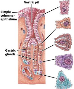
Gastric Gland Cell Types and Secretions
Surface mucous and mucous neck cells: Secrete mucus for protection
Parietal cells: Produce hydrochloric acid (HCl) and intrinsic factor (for vitamin B12 absorption)
Chief cells: Secrete pepsinogen (converted to pepsin by HCl) and gastric lipase
G cells: Endocrine cells that secrete gastrin (stimulates gastric activity)
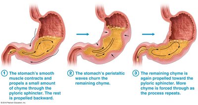
Digestion in the Stomach
Mechanical digestion: Mixing waves churn food into chyme
Chemical digestion: HCl denatures proteins, activates pepsinogen; pepsin digests proteins
Salivary amylase is inactivated by stomach acid
Regulation of Gastric Secretion and Motility
Cephalic phase: Triggered by sight, smell, taste, or thought of food; stimulates gastric secretion via vagus nerve (CN X)
Gastric phase: Initiated by food in the stomach; stretch and chemoreceptors increase secretion and motility
Intestinal phase: Begins as chyme enters the duodenum; enterogastric reflex and hormones (secretin, CCK, VIP) inhibit gastric activity
Small Intestine: Structure and Function
Gross Anatomy of the Small Intestine
~6 meters long; extends from pyloric sphincter to ileocecal valve
Subdivisions: Duodenum (retroperitoneal), jejunum, ileum (both attached by mesentery)
Major site of digestion and absorption
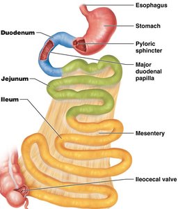
Duodenum, Jejunum, and Ileum
Duodenum: Shortest part (~25 cm), receives bile and pancreatic ducts, contains Brunner's glands (alkaline mucus)
Jejunum: ~2.5 m, main site of nutrient absorption
Ileum: ~3.6 m, joins large intestine at ileocecal valve, contains Peyer's patches (immune function)
Blood Supply and Innervation
Arterial supply: Superior mesenteric artery
Venous drainage: Superior mesenteric vein → hepatic portal vein → liver
Innervation: Vagus nerve (parasympathetic), thoracic splanchnic nerves (sympathetic)
Histology of the Small Intestine
Epithelium: Simple columnar (enterocytes) specialized for absorption; goblet cells secrete mucus
Surface area adaptations: Circular folds (plicae circulares), villi, microvilli (brush border)
Intestinal crypts: Secrete intestinal juice, contain enteroendocrine cells (hormones), Paneth cells (antimicrobial), stem cells
Digestion in the Small Intestine
Mechanical digestion: Segmentation and peristalsis mix and move chyme; bile aids fat digestion
Chemical digestion: Enzymes from pancreas and brush border complete digestion of carbohydrates, proteins, and lipids
Summary Table: Layers of the GI Tract
Layer | Main Components | Function |
|---|---|---|
Mucosa | Epithelium, lamina propria, muscularis mucosae | Secretion, absorption, protection |
Submucosa | Connective tissue, blood/lymph vessels, glands, submucosal plexus | Support, secretion regulation |
Muscularis externa | Smooth muscle (circular, longitudinal), myenteric plexus | Motility (peristalsis, segmentation) |
Serosa | Visceral peritoneum | Protection, reduces friction |
Key Equations and Concepts
Hydrolysis (general reaction for digestion):
Enzyme action (example: starch breakdown):
Additional info: The notes above integrate and expand upon the provided lecture content, ensuring a comprehensive, exam-ready summary for college-level Anatomy & Physiology students.
