 뒤로
뒤로Anatomy & Physiology: The Human Body - Orientation and Organization
스터디 가이드 - 스마트 노트
 자료에 맞춘 맞춤형 노트, 핵심 정의, 예시, 맥락을 확장해 제공합니다.
What is Anatomy and Physiology?
Definitions and Distinctions
Anatomy and physiology are two closely related fields that study the structure and function of the human body. Anatomy focuses on the physical structures, while physiology examines how those structures work.
Anatomy: The study of the body's structure, including organs, tissues, and cells.
Physiology: The study of the body's functions and processes, such as how organs operate and interact.
Principle of Complementarity: Structure and function are interdependent; understanding one requires knowledge of the other.
Example: The heart's anatomy (valves, chambers) determines its function (pumping blood).
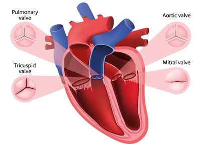
Additional info: The four main heart valves (pulmonary, aortic, tricuspid, mitral) ensure unidirectional blood flow, illustrating the relationship between structure and function.
Levels of Organization
Hierarchy of Biological Structure
The human body is organized into a hierarchy of levels, from the smallest chemical components to the entire organism.
Atomic and Molecular Level: Atoms and molecules form the basis of all biological structures.
Macromolecule Level: Large molecules such as proteins and DNA.
Cellular Level: Cells are the basic units of life.
Tissue Level: Groups of similar cells performing a common function.
Organ Level: Structures composed of multiple tissue types.
Organism Level: The complete living individual.
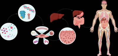
Example: Statins inhibit an enzyme at the molecular level, affecting cholesterol production at the organ level (liver), and ultimately impacting the whole organism.
Variation in Anatomy and Physiology
Anatomical Variation and Reference Bodies
There is significant variation in human anatomy and physiology. Reference bodies are used to standardize learning and communication.
Anatomical Variation: Differences in structure, such as blood vessel branching patterns.
Reference Body: A standardized model, typically a healthy young adult, used for teaching.
Normal Variation: Variations that do not affect function are considered normal.


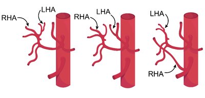
Example: The arrangement of hepatic arteries varies among individuals, but textbooks present a standard pattern for clarity.
Introduction to Organ Systems
Overview of Major Organ Systems
The human body is divided into organ systems, each responsible for specific functions. Systems are grouped by their primary roles.
Protection, Structure, & Support: Integumentary, skeletal, muscular systems.
Communication & Integration: Nervous and endocrine systems.
Transport & Immunity: Cardiovascular and lymphatic systems.
Nutrient, Gas, & Waste Exchange: Respiratory, digestive, urinary systems.
Reproduction: Male and female reproductive systems.
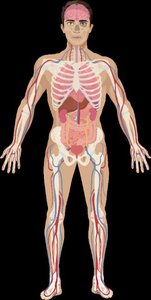
Additional info: Each system is composed of organs that work together to maintain homeostasis and support life.
Anatomical Position
Standard Reference for Describing the Body
The anatomical position is a universally accepted reference posture for describing locations and directions in the body.
Body upright, face forward, feet slightly apart, arms at sides, palms facing forward.
Left and right refer to the subject's left and right.
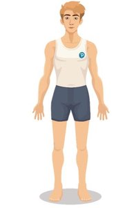
Importance: Ensures clear communication and minimizes errors in medical and anatomical descriptions.
Directional Terms
Describing Locations and Relationships
Directional terms are used to compare the locations of structures within the body, always referencing the anatomical position.
Superior: Toward the head.
Inferior: Toward the feet.
Anterior (Ventral): Toward the front.
Posterior (Dorsal): Toward the back.
Medial: Toward the midline.
Lateral: Away from the midline.
Proximal: Closer to the point of attachment (limbs).
Distal: Farther from the point of attachment (limbs).
Superficial: Closer to the surface.
Deep: Further from the surface.
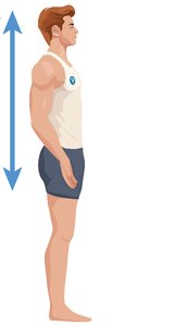
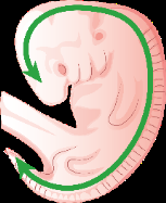
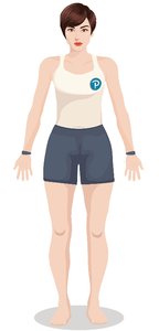
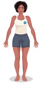
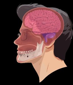
Additional info: Directional terms are essential for describing the precise location of injuries, procedures, or anatomical features.
Anatomical Terms for Body Regions
Regional Terminology
Specific terms are used to describe regions of the body, often derived from Greek or Latin.
Head and Neck: Cephalic, cervical, orbital, nasal, buccal, otic, mental, occipital, frontal.
Trunk: Thoracic, abdominal, pelvic, umbilical, inguinal, pubic, axillary, sternal.
Back: Scapular, lumbar, sacral, perineal, vertebral, gluteal, olecranal.
Arm and Hand: Brachial, antebrachial, acromial, axillary, carpal, pollex, digital, manus, antecubital.
Leg and Foot: Femoral, coxal, patellar, popliteal, sural, crural, tarsal, hallux, plantar, pedal, metatarsal, calcaneal.
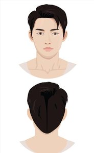

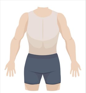
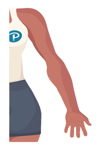
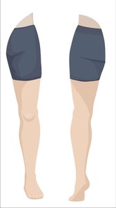
Additional info: Mastery of regional terms is crucial for accurate communication in clinical and academic settings.
Abdominopelvic Quadrants and Regions
Divisions of the Abdomen and Pelvis
The abdomen and pelvis are divided into quadrants and regions for clinical and anatomical reference.
Quadrants: Right Upper (RUQ), Left Upper (LUQ), Right Lower (RLQ), Left Lower (LLQ).
Regions: Right/Left hypochondriac, epigastric, right/left lumbar, umbilical, right/left inguinal, hypogastric.


Example: Tenderness in the RLQ may indicate appendicitis; the LUQ contains the stomach and spleen.
Anatomical Planes and Sections
Planes Used for Dissection and Imaging
Anatomical planes are imaginary lines used to divide the body for study, imaging, or surgery.
Frontal (Coronal) Plane: Divides anterior and posterior.
Sagittal Plane: Divides left and right; midsagittal is at the midline, parasagittal is off-center.
Transverse Plane: Divides superior and inferior.
Oblique Plane: Divides at an angle.
Additional info: Sections are created by cuts along these planes, useful in imaging techniques like MRI and CT scans.
Organization of the Body: Body Cavities
Major Body Cavities and Their Contents
The body contains cavities that house organs and provide protection and compartmentalization.
Anterior (Ventral) Cavity: Contains thoracic and abdominopelvic cavities.
Posterior (Dorsal) Cavity: Contains cranial and vertebral cavities.
Thoracic Cavity: Houses heart and lungs, protected by rib cage.
Abdominopelvic Cavity: Houses digestive, urinary, and reproductive organs.
Example: The brain is in the cranial cavity; the stomach is in the abdominal cavity.
Serous Membranes
Structure and Function of Serous Membranes
Serous membranes are double-layered tissues that line body cavities and cover organs, providing lubrication and protection.
Visceral Layer: Attached to the organ.
Parietal Layer: Attached to the cavity wall.
Serous Cavity: Space between layers, filled with serous fluid.
Serous Fluid: Reduces friction between organs and cavity walls.
Major Serous Membranes:
Pleura: Surrounds lungs.
Pericardium: Surrounds heart.
Peritoneum: Surrounds abdominal organs.
Example: Inflammation of the pericardium (pericarditis) causes chest pain due to reduced lubrication.
Additional info: Serous membranes are essential for organ movement and protection, and their dysfunction can lead to disease.
