 뒤로
뒤로Blood: Leukocytes, Platelets, and Hemostasis (Chapter 16, Part B)
스터디 가이드 - 스마트 노트
 자료에 맞춘 맞춤형 노트, 핵심 정의, 예시, 맥락을 확장해 제공합니다.
Leukocytes (White Blood Cells)
General Structure and Functional Characteristics
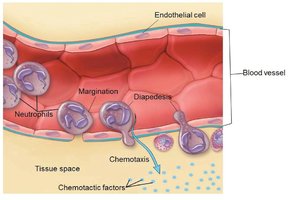
Leukocytes, or white blood cells (WBCs), are the only complete cells in blood, containing nuclei and organelles. They play a crucial role in defending the body against pathogens and foreign substances. Leukocytes can leave capillaries by a process called diapedesis, move through tissue spaces using amoeboid motion, and are attracted to sites of infection or injury by positive chemotaxis.
Types and Relative Percentages of Leukocytes
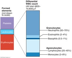
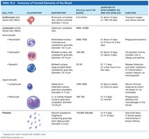
Leukocytes are classified into two main groups based on the presence or absence of cytoplasmic granules: granulocytes and agranulocytes. The differential WBC count provides the relative percentages of each type in normal blood.
Granulocytes
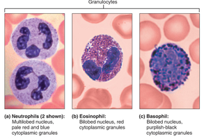
Granulocytes include neutrophils, eosinophils, and basophils. These cells have lobed nuclei and visible cytoplasmic granules containing enzymes that destroy pathogens.
Neutrophils
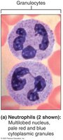
Neutrophils are the most numerous WBCs, accounting for about 50–70% of all leukocytes. They are the body's first line of defense and are highly phagocytic, especially against bacteria. Their granules contain hydrolytic enzymes, lysozyme, and antimicrobial proteins called defensins.
Eosinophils
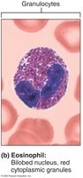
Eosinophils have a bilobed nucleus and red cytoplasmic granules. They increase in number during parasitic infections and are involved in immune responses, especially in allergic and inflammatory reactions. Eosinophils release enzymes that target the surface of parasitic worms and chemical mediators that modulate inflammation.
Basophils
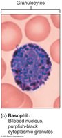
Basophils are the rarest leukocytes (0.5–1%). They have large, purplish-black granules containing histamine, a vasodilator that attracts other WBCs to inflamed sites. Basophils are functionally similar to mast cells and play a role in allergic responses.
Agranulocytes
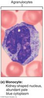
Agranulocytes lack visible cytoplasmic granules and include lymphocytes and monocytes. Their nuclei are typically spherical or kidney-shaped.
Lymphocytes
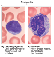
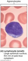
Lymphocytes make up about 25% of WBCs and are mostly found in lymphoid tissues such as lymph nodes and the spleen. They are crucial for immunity and are divided into two main types:
T lymphocytes (T cells): Act against virally infected cells and tumor cells.
B lymphocytes (B cells): Differentiate into plasma cells that produce antibodies.
Monocytes
Monocytes are the largest leukocytes (3–8% of WBCs) with U- or kidney-shaped nuclei. They leave the bloodstream, enter tissues, and differentiate into macrophages, which are actively phagocytic and crucial in defense against viruses, intracellular bacterial parasites, and chronic infections. Monocytes also activate lymphocytes to mount an immune response.
Leukopoiesis – WBC Production
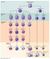
Leukopoiesis is the process of WBC production, occurring in the red bone marrow. It is regulated by chemical messengers such as interleukins and colony-stimulating factors (CSFs). T lymphocyte precursors mature in the thymus, while B lymphocyte precursors mature in the bone marrow.
Leukocyte Disorders
Leukemias: Cancers involving overproduction of abnormal WBCs.
Infectious mononucleosis: A highly contagious viral disease caused by the Epstein-Barr virus, often seen in young adults.
Platelets
Structure and Function
Platelets are cell fragments derived from megakaryocytes. Their granules contain chemicals essential for the clotting process. Platelets form a temporary plug to help seal breaks in blood vessels. In circulation, platelets are kept inactive by nitric oxide and prostacyclin released by endothelial cells.
Formation of Platelets
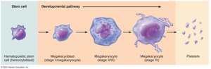
Platelet production is regulated by the hormone thrombopoietin. Megakaryocytes undergo mitosis without cytokinesis, resulting in a large cell with a multilobed nucleus. Cytoplasmic projections break off into the capillary lumen, forming platelets. Platelets have a lifespan of about 10 days.
Summary of Formed Elements
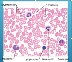
The formed elements of blood include erythrocytes, leukocytes, and platelets. Each type has distinct structural and functional characteristics.
Cell Type | Description | Function |
|---|---|---|
Erythrocytes | Biconcave, anucleate | Transport oxygen and carbon dioxide |
Neutrophils | Multilobed nucleus, pale granules | Phagocytize bacteria |
Eosinophils | Bilobed nucleus, red granules | Kill parasitic worms, modulate allergies |
Basophils | Bilobed nucleus, purplish-black granules | Release histamine, mediate inflammation |
Lymphocytes | Large nucleus, thin rim of cytoplasm | Immune response via antibodies or direct attack |
Monocytes | Kidney-shaped nucleus, abundant cytoplasm | Phagocytosis, develop into macrophages |
Platelets | Cell fragments | Seal small tears in blood vessels, clotting |
Hemostasis – Stopping Bleeding
Hemostasis is the process that stops bleeding and involves three major steps:
Step 1: Vascular spasm
Step 2: Platelet plug formation
Step 3: Coagulation (blood clotting)
Step 1: Vascular Spasm
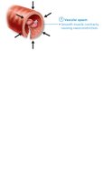
Vascular spasm is the immediate constriction of a damaged blood vessel, triggered by direct injury to vascular smooth muscle and chemicals released by damaged endothelial cells. This reduces blood flow and limits blood loss.
Step 2: Platelet Plug Formation
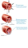
When a blood vessel is damaged, collagen fibers are exposed, and platelets adhere to them. Activated platelets release chemicals such as ADP (which attracts more platelets), serotonin, and thromboxane A2 (which enhance spasm and aggregation), creating a positive feedback cycle that forms a platelet plug.
Step 3: Coagulation (Blood Clotting)
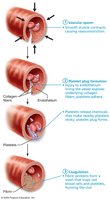
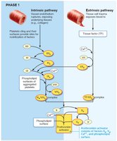
Coagulation reinforces the platelet plug with fibrin threads, transforming blood from a liquid to a gel. This process involves a cascade of reactions using clotting factors (procoagulants), many of which require vitamin K. Coagulation occurs in three phases:
Phase 1: Two pathways (intrinsic and extrinsic) lead to the formation of prothrombin activator.
Phase 2: Prothrombin activator converts prothrombin to thrombin.
Phase 3: Thrombin converts fibrinogen to fibrin, forming the structural basis of the clot. Factor XIII stabilizes the clot.
Clot Retraction and Vessel Repair
Clot retraction stabilizes the clot as actin and myosin in platelets contract, pulling on fibrin strands and squeezing serum from the clot. This draws the edges of the ruptured vessel together. Platelet-derived growth factor (PDGF) stimulates fibroblasts and smooth muscle cells to rebuild the vessel wall, while vascular endothelial growth factor (VEGF) restores the endothelial lining.
Fibrinolysis – Removal of Clots
Fibrinolysis is the process of removing clots after vessel repair. Plasminogen, trapped in the clot, is converted to plasmin by tissue plasminogen activator (tPA). Plasmin digests fibrin, dissolving the clot.
Blood Clotting Factors (Procoagulants)
Blood clotting involves a series of factors, each with a specific role in the coagulation cascade. These are summarized in the following table:
Factor | Name | Function |
|---|---|---|
I | Fibrinogen | Converted to fibrin by thrombin |
II | Prothrombin | Converted to thrombin |
III | Tissue factor (TF) | Activates extrinsic pathway |
IV | Calcium ions (Ca2+) | Required for many steps in coagulation |
V-XIII | Various plasma proteins | Act as enzymes or cofactors in the cascade |
Additional info: For a complete list of clotting factors, refer to Table 16.3 in your textbook.
