 뒤로
뒤로Blood: Plasma Proteins, Erythropoiesis, Platelets, and Leukocytes
스터디 가이드 - 스마트 노트
 자료에 맞춘 맞춤형 노트, 핵심 정의, 예시, 맥락을 확장해 제공합니다.
Blood Composition and Plasma Proteins
Overview of Blood and Plasma Proteins
Blood is a specialized connective tissue composed of plasma and formed elements. Plasma proteins play critical roles in maintaining homeostasis, transporting substances, and defending the body against pathogens.
Albumin: Synthesized in the liver, albumin maintains blood colloid osmotic pressure, helps retain fluid in the bloodstream, and transports substances such as fatty acids, hormones, drugs, and bilirubin.
Globulins (α, β, γ): α and β globulins are produced in the liver, while γ globulins are produced by plasma cells/lymphoid tissues. They transport lipids, metal ions, and vitamins, contribute to immunity, and γ globulins function as antibodies.
Fibrinogen: Produced in the liver, fibrinogen is essential for blood clotting and is converted to fibrin during coagulation to form blood clots.
Plasma Protein | Site of Synthesis | Main Functions |
|---|---|---|
Albumin | Liver | Maintains blood colloid (oncotic) osmotic pressure, helps keep fluid in the bloodstream, and transports substances such as fatty acids, hormones, drugs, and bilirubin. |
Globulins (α, β, γ) | α and β globulins: Liver; γ globulins: Plasma cells/lymphoid tissues | Transport lipids, metal ions, and vitamins; contribute to immunity; gamma globulins function as antibodies that help defend against infection. |
Fibrinogen | Liver | Essential for blood clotting; converted to fibrin during coagulation to form blood clots. |
Erythropoiesis and Erythropoietin (EPO)
Regulation of Red Blood Cell Production
Erythropoiesis is the process of red blood cell (RBC) production, primarily regulated by the hormone erythropoietin (EPO). EPO ensures adequate oxygen delivery by stimulating RBC formation in response to hypoxia.
Site of EPO Production: Primarily the kidneys (renal cells); a small amount is produced by the liver.
Stimulus for EPO Release: Low blood oxygen levels (hypoxia), such as during anemia, blood loss, high altitude exposure, or reduced oxygen delivery to tissues.
Target Tissue(s): Red bone marrow, specifically erythroid progenitor and precursor cells that develop into red blood cells.
Action of EPO: Stimulates erythropoiesis by promoting the survival, proliferation, and differentiation of erythroid precursor cells, increasing the number of circulating RBCs and improving oxygen-carrying capacity.
Item | Answer |
|---|---|
Site of EPO Production | Primarily the kidneys (renal cells). A small amount is produced by the liver. |
Stimulus for EPO Release | Low blood oxygen levels (hypoxia), such as during anemia, blood loss, high altitude exposure, or reduced oxygen delivery to tissues. |
Target Tissue(s) | Red bone marrow, specifically erythroid progenitor and precursor cells that develop into red blood cells. |
Action of EPO | Stimulates erythropoiesis (red blood cell production) by promoting the survival, proliferation, and differentiation of erythroid precursor cells, leading to an increased number of circulating red blood cells and improved oxygen-carrying capacity of the blood. |
Platelets and Thrombopoiesis
Formation and Function of Platelets
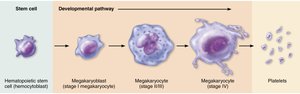
Platelets, or thrombocytes, are small cell fragments essential for blood clotting. They are produced through the process of thrombopoiesis, regulated by the hormone thrombopoietin (TPO).
Origin: Platelets are derived from megakaryocytes, which develop from hematopoietic stem cells in the bone marrow.
Developmental Pathway: Hematopoietic stem cell → Megakaryoblast → Megakaryocyte (stages I-IV) → Platelets (by cytoplasmic fragmentation).
Function: Platelets help stop bleeding by forming plugs in blood vessel injuries and releasing factors that promote clotting.
Leukocytes (White Blood Cells): Types and Functions
Classification and Roles of Leukocytes
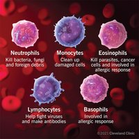
Leukocytes are the body's primary defense against infection and disease. They are classified into granulocytes and agranulocytes based on the presence or absence of cytoplasmic granules.
Granulocytes: Neutrophils, eosinophils, basophils
Agranulocytes: Lymphocytes, monocytes
Relative Abundance and Blood Smear Appearance
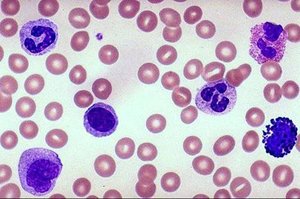
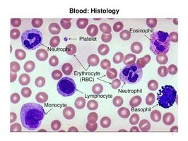
The five types of leukocytes vary in abundance and can be identified in blood smears by their distinct morphology and staining characteristics.
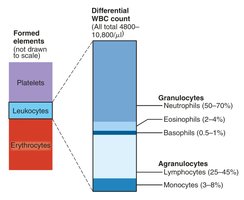
Neutrophils: 50–70% of WBCs; multi-lobed nucleus, pale granules; phagocytize bacteria.
Lymphocytes: 25–45%; large, round nucleus; involved in immune responses (T and B cells).
Monocytes: 3–8%; kidney-shaped nucleus; differentiate into macrophages.
Eosinophils: 2–4%; bi-lobed nucleus, red granules; combat parasites, involved in allergies.
Basophils: 0.5–1%; U/S-shaped nucleus, dark granules; release histamine in inflammation.
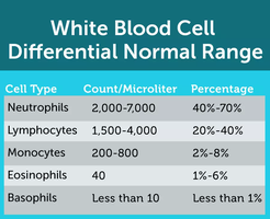
Normal Cell Counts of WBCs in Adults
Normal ranges for each type of white blood cell are important for diagnosing various conditions.
Cell Type | Count/Microliter | Percentage |
|---|---|---|
Neutrophils | 2,000–7,000 | 40%–70% |
Lymphocytes | 1,500–4,000 | 20%–40% |
Monocytes | 200–800 | 2%–8% |
Eosinophils | 40 | 1%–6% |
Basophils | Less than 10 | Less than 1% |
Summary Table: Leukocyte Types and Functions
Leukocyte | Main Function |
|---|---|
Neutrophils | Kill bacteria, fungi, and foreign debris |
Monocytes | Clean up damaged cells |
Eosinophils | Kill parasites, cancer cells, and involved in allergic response |
Lymphocytes | Help fight viruses and make antibodies |
Basophils | Involved in allergic response |
Blood Smear Histology
Microscopic Identification of Blood Cells
Blood smears stained with Wright's stain allow for the identification of different blood cells based on their morphology and staining properties. Neutrophils, eosinophils, basophils, lymphocytes, and monocytes can be distinguished by their nuclear shape and cytoplasmic granules.
Additional info:
Granulocytes are characterized by the presence of cytoplasmic granules and include neutrophils, eosinophils, and basophils. Agranulocytes lack visible granules and include lymphocytes and monocytes.
Platelets are not true cells but fragments of megakaryocytes, essential for hemostasis.
EPO is a key hormone in the regulation of erythropoiesis, especially in response to hypoxic conditions.
